
Maxillary Palp Collection
The intricate world of insect anatomy never ceases to amaze. Take a closer look at the fascinating maxillary palp, a sensory organ found in various insects


All Professionally Made to Order for Quick Shipping
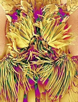
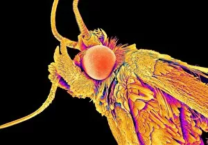
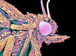
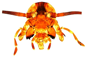
The intricate world of insect anatomy never ceases to amaze. Take a closer look at the fascinating maxillary palp, a sensory organ found in various insects. In these captivating scanning electron microscope (SEM) images, we delve into the hidden details of different species. Starting with the moth heads captured under SEM C015 / 8073 and SEM C015 / 8074, we witness the delicate structure of their maxillary palps. These elongated appendages are adorned with tiny sensilla that enable moths to detect chemical cues in their environment, aiding them in finding mates or locating food sources. Moving on to SEM C015 / 8071 and SEM C015 / 8072, another set of moth heads reveals further intricacies within their maxillary palps. The fine hairs and sensory receptors present on these organs showcase nature's remarkable adaptation for survival. Next up are striking images of geometer moths observed under SEM. Their maxillary palps exhibit unique variations compared to those of moths seen earlier. These adaptations reflect diverse ecological roles played by different species within this family. Shifting our focus towards ant heads examined through SEM, we uncover yet another intriguing aspect – ants possess maxillary palps too. These minuscule but essential structures aid ants in detecting pheromones released by their colony members for communication purposes. Concluding our exploration is one last glimpse into a moth head captured under an SEM lens. This image serves as a reminder that even within a single insect order like Lepidoptera (moths), there exists incredible diversity when it comes to the structure and function of their maxillary palps. These mesmerizing micrographs offer us valuable insights into the complexity and beauty inherent in nature's smallest creations. They remind us that every organism has its own story written within its anatomy – waiting patiently for curious minds like ours to unravel its secrets.