
Lymph Node Collection
The lymph node: a vital player in our body's defense system. 🌟 Step into the intricate world of the human lymphatic system with this captivating illustration


All Professionally Made to Order for Quick Shipping
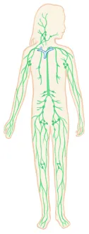
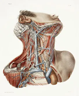
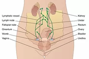
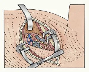
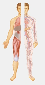
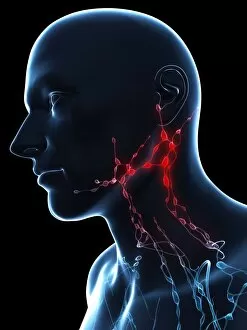
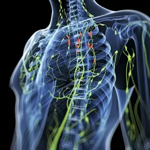
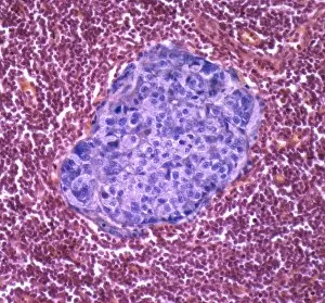
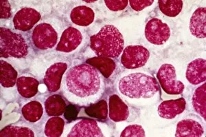
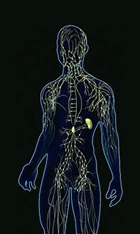
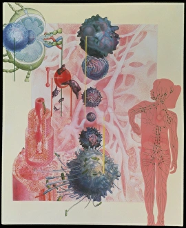
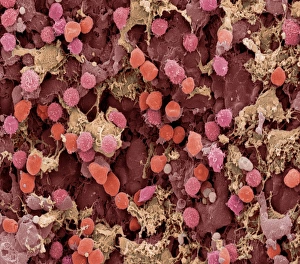
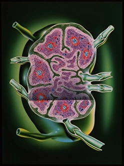
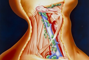
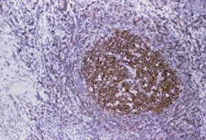
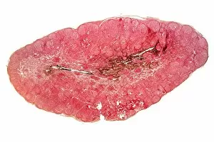
The lymph node: a vital player in our body's defense system. 🌟 Step into the intricate world of the human lymphatic system with this captivating illustration. 🧪 From neck anatomy to 19th-century artwork, each detail brings us closer to understanding these remarkable nodes. Peering through an abdominal incision held open by spreaders, we witness the delicate dance between human lungs and blood vessels. This mesmerizing image reveals just how interconnected our internal systems truly are. Zooming in further, we encounter macrophages and lymphocytes under a TEM microscope. These tiny warriors tirelessly patrol our bodies, ready to combat any foreign invaders that may threaten our health. Intriguing female abdominal anatomy captured in stunning artwork showcases the significance of lymph nodes within our bodies' natural rhythm. They serve as guardians against potential threats, ensuring harmony within. A digital illustration exposes a tumor nestled within a lymph node – a stark reminder of the challenges some face on their medical journey. It highlights the importance of early detection and treatment options available today. Cross-section illustrations grant us an intimate look at both healthy and cancerous nodes requiring lumpectomy or mastectomy procedures for breast cancer patients. These visuals shed light on medical advancements aimed at saving lives every day. As we explore further, an intricately detailed diagram maps out the entire lymphatic system – its conducting pathways intertwined with numerous essential nodes throughout our body's landscape. This comprehensive illustration serves as a testament to how crucial these small but mighty structures are for maintaining optimal health and well-being. The lymph node is not merely another part of our anatomy; it is an unsung hero silently working behind-the-scenes to protect us from harm.