
Luxation Collection
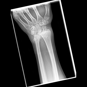
Luxation, also known as dislocation, is a condition that occurs when the bones in a joint are forced out of their normal position


All Professionally Made to Order for Quick Shipping
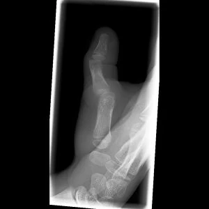
Luxation, also known as dislocation, is a condition that occurs when the bones in a joint are forced out of their normal position. One common type of the "luxation sous coracoidienne de l'épaule, " which refers to the displacement of the head of the humerus bone in the shoulder joint. This condition can cause severe pain and limited mobility. Injuries such as a broken elbow or fractured ankle often accompany luxations. X-ray images like C017 / 7266, C017 / 7565, C017 / 7183, and C017 / 7563 reveal these dislocations clearly. These images show how bones can be forcefully moved from their usual alignment due to accidents or trauma. The consequences of they can vary depending on the severity and location of the injury. For instance, lower back pain may result from a luxated spine depicted in conceptual artwork illustrating this discomfort. X-rays like C017 / 7979 and C017 / 7968 display broken elbows caused by luxations while others like C017 / 7978 demonstrate pinned fractures or dislocations in ankles. These images highlight both the damage done to bones during an injury and medical interventions used for treatment. Dislocated thumbs are another example where X-ray imaging (C017/7805) helps diagnose this specific type accurately.