Fraud Blocker Skip to main content

Luxated Collection

"Capturing the Fragility of Bones: A Glimpse into Luxated Injuries" In the world of X-rays

Background imageLuxated Collection: Broken elbow, X-ray C017 / 7266

Broken elbow, X-ray C017 / 7266
Broken elbow. X-ray of the elbow of a 4 year old female with a displaced supracondylar fracture. The fracture is at the base of the humerus, the upper arm bone (upper frame)

Background imageLuxated Collection: Dislocated wrist, X-ray C017 / 7565

Dislocated wrist, X-ray C017 / 7565
Dislocated wrist. X-ray of the wrist of a 16 year old male that has been dislocated

Background imageLuxated Collection: Dislocated finger, X-ray C017 / 7183

Dislocated finger, X-ray C017 / 7183
Dislocated finger. X-ray of the left hand of a 51 year old female patient with a dislocated middle finger. Once the bones have been manipulated back into place treatment is with immobilisation

Background imageLuxated Collection: Dislocated wrist, X-ray C017 / 7563

Dislocated wrist, X-ray C017 / 7563
Dislocated wrist. X-ray of the wrist of a 16 year old male that has been dislocated

Background imageLuxated Collection: Broken elbow, X-ray C017 / 7979

Broken elbow, X-ray C017 / 7979
Broken elbow. X-ray of the elbow of a 4 year old female with a displaced supracondylar fracture. The fracture is at the base of the humerus, the upper arm bone (upper frame)

Background imageLuxated Collection: Pinned broken ankle, X-ray C017 / 7978

Pinned broken ankle, X-ray C017 / 7978
Pinned broken ankle. X-ray of the ankle of a 32 year old male that has been pinned after a parachuting accident. The ankle was dislocated between the tibia (shin bone)

Background imageLuxated Collection: Fractured and dislocated ankle, X-ray C017 / 7976

Fractured and dislocated ankle, X-ray C017 / 7976
Fractured and dislocated ankle. X-ray of the ankle of a 32 year old male who has been in a parachuting accident. The ankle is dislocated between the tibia (shin bone)

Background imageLuxated Collection: Broken elbow, X-ray C017 / 7968

Broken elbow, X-ray C017 / 7968
Broken elbow. X-ray of the elbow of a 5 year old female with a displaced supracondylar fracture. The fracture is at the base of the humerus, the upper arm bone (upper frame)

Background imageLuxated Collection: Pinned fractured elbow, X-ray C017 / 7969

Pinned fractured elbow, X-ray C017 / 7969
Pinned fractured elbow. X-ray of the elbow of a 5 year old female that has been internally fixed after a displaced supracondylar fracture

Background imageLuxated Collection: Dislocated thumb, X-ray C017 / 7805

Dislocated thumb, X-ray C017 / 7805
Dislocated thumb. X-ray of the right hand of a 10 year old male patient with a dislocated thumb

Background imageLuxated Collection: Dislocated fingers, X-ray C017 / 7754

Dislocated fingers, X-ray C017 / 7754
Dislocated fingers. X-ray of the hand of a 60 year old male patient who has dislocated his third, fourth and fifth fingers

Background imageLuxated Collection: Dislocated toe, X-ray

Dislocated toe, X-ray
Dislocated toe. Coloured X-ray of the foot of a 49 year old woman with a dislocated toe. The dislocated joint is shown in yellow. It is the fourth proximal interphalangeal joint


All Professionally Made to Order for Quick Shipping

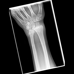
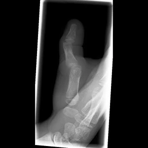
"Capturing the Fragility of Bones: A Glimpse into Luxated Injuries" In the world of X-rays, a series of haunting images reveal the delicate nature of our skeletal framework. Amongst them, an array of fractures and dislocations paint a vivid picture of human vulnerability. The first glimpse takes us to a broken elbow, its jagged lines etched across X-ray C017 / 7266. The intricate network of shattered bones serves as a stark reminder that even seemingly sturdy joints can succumb to immense pressure. Moving along, we encounter another dislocated wrist in X-ray C017 / 7565. The once harmonious alignment now disrupted, leaving behind pain and instability in its wake, and is here that we realize how easily life's unexpected twists can lead to profound consequences. A dislocated finger captured in X-ray C017 / 7183 further emphasizes this fragility. What was once an agile digit becomes contorted and disjointed, reminding us that even the smallest appendages are not immune to injury's grasp. Continuing on our journey through these visual narratives, we stumble upon yet another dislocated wrist showcased in X-ray C017 / 7563. Its presence reinforces the notion that accidents often strike with repetitive force – relentless in their pursuit to disrupt our equilibrium. But it is not just wrists and elbows bearing witness to such misfortune; ankles too fall victim to bone-related woes. A pinned broken ankle stares back at us from X-ray C017 / 7978 - held together by metal reinforcements amidst a sea of fractures. This image stands testament to both medical intervention and resilience against adversity. Further down this path lies an ankle fractured and dislocated simultaneously (X-ray C017 / 7976). Here lies evidence that sometimes injuries overlap - causing unimaginable pain while challenging medical professionals' expertise as they navigate complex cases like these.