Fraud Blocker Skip to main content

Light Micrograph Collection (page 3)

Capturing the intricate beauty of biological structures, a light micrograph reveals the mesmerizing complexity of cerebellum tissue

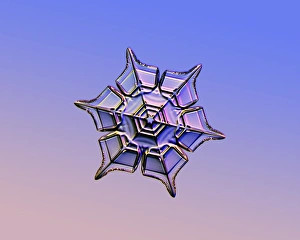
Background imageLight Micrograph Collection: Snowflake, light micrograph

Snowflake, light micrograph
Snowflake. Light micrograph of a stellar plate snowflake. Snowflakes are symmetrical ice crystals that form inside clouds when water vapour condenses directly into ice

Background imageLight Micrograph Collection: Gout crystals

Gout crystals. Polarised light micrograph of uric acid crystals in the synovial fluid (a lubricating liquid in joints) from a patient with gout

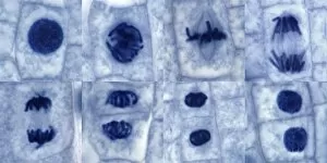
Background imageLight Micrograph Collection: Plant cell mitosis, light micrograph

Plant cell mitosis, light micrograph
Plant cell mitosis. Light micrograph of root tip cells from an onion (Allium sp.) undergoing cell division (mitosis). From top left to bottom right

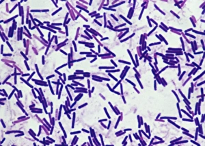
Background imageLight Micrograph Collection: Botulism bacteria

Botulism bacteria. Light micrograph of botulism bacteria (Clostridium botulinum). These bacteria occur naturally in soil. They produce botulinum toxin

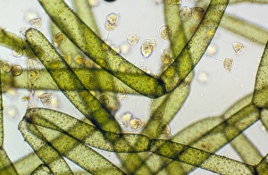
Background imageLight Micrograph Collection: LM of Vorticella ciliates on a green alga

LM of Vorticella ciliates on a green alga
Light micrograph of a group of Vorticella (small tethered balloons) adhering to the freshwater green alga Hydrodictyon (large green cells)

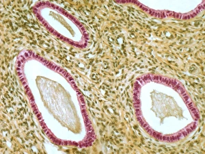
Background imageLight Micrograph Collection: Cervix, light micrograph F006 / 9805

Cervix, light micrograph F006 / 9805
Cervix. Light micrograph of a section though the mucosa of the cervix, the neck of the uterus. The purple-lined areas are cervical glands, which produce mucus (pink inside the glands)

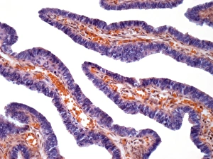
Background imageLight Micrograph Collection: Fallopian tube, light micrograph F006 / 9799

Fallopian tube, light micrograph F006 / 9799
Fallopian tube. Light micrograph of a cross-section through a fallopian tube. The lumen (white) is seen, along with mucosal folds lined with simple columnar ciliated epithelium (purple)

Background imageLight Micrograph Collection: Thymus gland, light micrograph C015 / 4970

Thymus gland, light micrograph C015 / 4970
Thymus gland. Light micrograph of a section through tissue from the thymus gland, part of the lymphoid system. The dense areas are the thymic cortex, which produces lots of lymphocytes

Background imageLight Micrograph Collection: Myeloblast blood cell, light micrograph

Myeloblast blood cell, light micrograph
Myeloblast blood cell. Light micrograph of blood cells, including a myeloblast, a precursor for a type of white blood cell (leucocyte). Myeloblasts differentiate into granulocytes

Background imageLight Micrograph Collection: Promyelocyte blood cell, light micrograph

Promyelocyte blood cell, light micrograph. This blood cell (centre) is a precursor for a type of white blood cell called a granulocyte, formed by granulopoiesis in the bone marrow

Background imageLight Micrograph Collection: Chronic lymphocytic leukaemia, micrograph

Chronic lymphocytic leukaemia, micrograph
Chronic lymphocytic leukaemia. Light micrograph of blood cells sampled from a lymphatic ganglion in a case of chronic lymphocytic leukaemia, also called chronic lymphoid leukaemia (CLL)

Background imageLight Micrograph Collection: Lancet liver fluke C014 / 4846

Lancet liver fluke C014 / 4846
Lancet liver fluke (Dicrocoelium dendriticum). This parasitic trematode (flatworm) tends to live in cattle or other grazing mammals

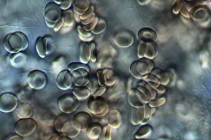
Background imageLight Micrograph Collection: Red blood cells, light micrograph C016 / 3035

Red blood cells, light micrograph C016 / 3035
Red blood cells. Differential interference contrast (DIC) micrograph of red blood cells (erythrocytes). Red blood cells are biconcave, giving them a large surface area for gas exchange

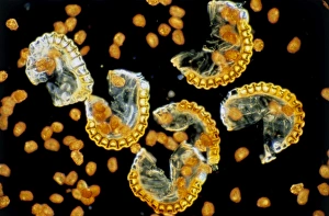
Background imageLight Micrograph Collection: Sporangia and spores of the male fern

Sporangia and spores of the male fern
Light micrograph of the sporangia (spore sacs) and spores of the male fern", Dryopteris filix-mas. The sporangia of Dryopteris are borne on the undersides of the leaf fronds in brown kidney

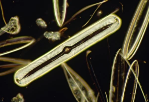
Background imageLight Micrograph Collection: LM of the diatom Pinnularia nobilis

LM of the diatom Pinnularia nobilis
Light micrograph of the diatom Pinnularia nobilis (centre), surrounded by other assorted diatoms. The diatoms are a distinctive group of single- celled algae containing about 10, 000 species

Background imageLight Micrograph Collection: Light Micrograph (SEM): Micro-filarial worm - Magnification x 3000 (if print A4 size: 29. 7 cm wide)

Light Micrograph (SEM): Micro-filarial worm - Magnification x 3000 (if print A4 size: 29. 7 cm wide)
LRDS-314 Light Micrograph (SEM): Micro-filarial worm Magnification x 3000 (if print A4 size: 29.7 cm wide) Wuchereria bancrofti Elephantiasis David Spears (Last Refuge)

Background imageLight Micrograph Collection: Wuchereria bancrofti parasite

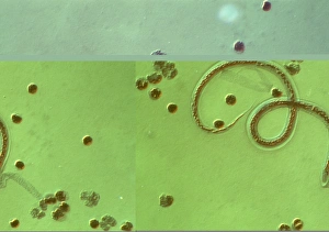
Wuchereria bancrofti parasite
Wuchereria bancrofti. Light micrograph of the microfilaria larval stage of the parasitic worm Wuchereria bancrofti, which causes filariasis in humans. W

Background imageLight Micrograph Collection: C. elegans worm

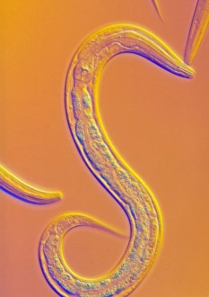
C. elegans worm
Caenorhabditis elegans nematode worm, light micrograph. This soil-dwelling hermaphrodite nematode worm is one of the most studied animals in biological and genetic research

Background imageLight Micrograph Collection: LM of the marine dinoflagellate, Ceratium sp

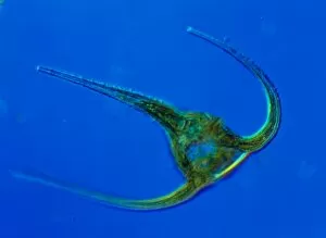
LM of the marine dinoflagellate, Ceratium sp
Dinoflagellate. Light micrograph of the marine dinoflagellate Ceratium. Found in both freshwater and saltwater, dinoflagellates are single-celled protozoa that float in the water column as plankton

Background imageLight Micrograph Collection: Euglena gracilis

Euglena gracilis protists. Light micrograph of a group of Euglena gracilis protists. These organisms possess a unique combination of plant and animal characters. Like the algae, E

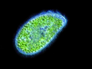
Background imageLight Micrograph Collection: Paramecium bursaria protozoan, micrograph

Paramecium bursaria protozoan, micrograph
Paramecium bursaria protozoan, light micrograph. This ciliate protozoan inhabits freshwater, where it feeds mainly on bacteria

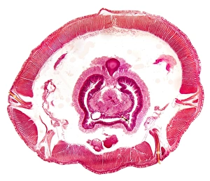
Background imageLight Micrograph Collection: Earthworm, transverse section

Earthworm, transverse section
Earthworm. Light micrograph of a transverse section through the body of a round segmented earthworm (Lumbricus terrestris) in the intestinal region

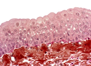
Background imageLight Micrograph Collection: Bladder epithelium, light micrograph

Bladder epithelium, light micrograph
Bladder epithelium. Light micrograph of a vertical section through the wall of the urinary bladder. The inner surface is at top

Background imageLight Micrograph Collection: Lung alveoli

Lung alveoli. Light micrograph of a section through normal human lung tissue showing alveoli. These are tiny air sacs where most of the diffusion of gases to and from the blood occurs

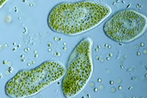
Background imageLight Micrograph Collection: Paramecium protozoa, light micrograph

Paramecium protozoa, light micrograph
Paramecium bursaria protozoa, light micrograph. These ciliate protozoa inhabit freshwater, where they feed mainly on bacteria

Background imageLight Micrograph Collection: Whale bone tissue, light micrograph

Whale bone tissue, light micrograph

Background imageLight Micrograph Collection: Hazel nut, light micrograph

Hazel nut, light micrograph
Hazel nut. Light micrograph of a section through a nut from a common hazel (Corylus avellana) tree, showing the large amount of fat (red) stored within endosperm tissue

Background imageLight Micrograph Collection: Trichodina parasite, light micrograph

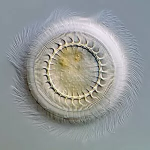
Trichodina parasite, light micrograph
Trichodina parasite. Differential interference contrast micrograph of Trichodina pediculus. This ciliate parasite lives on the skin, gills and mucous membranes of many aquatic animals

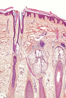
Background imageLight Micrograph Collection: Skin and hair follicles, light micrograph

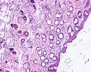
Skin and hair follicles, light micrograph
Skin and hair follicles. Light micrograph of a section through skin, showing hair follicles (dark purple circular structures). Haematoxylin and eosin stain

Background imageLight Micrograph Collection: Iris root, light micrograph

Iris root, light micrograph
Iris root. Light micrograph of a section through the root of an Iris plant (Iris germanica) showing a vascular cylinder. The cylinder is comprised of a central cluster of parenchyma cells (red)

Background imageLight Micrograph Collection: Light Micrograph : Flattened Mayfly Nymph, Magnification x 10 (A4 size: 29. 7 cm width)

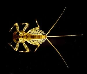
Light Micrograph : Flattened Mayfly Nymph, Magnification x 10 (A4 size: 29. 7 cm width)
LRDS-80 Flattened Mayfly Nymph Light Micrograph Ecdyonurus venosus Magnification x 10 (A4 size: 29.7 cm width) Credit: David Spears (last refuge)

Background imageLight Micrograph Collection: Light Micrograph (LM): Transverse section of Dehiscence Lilium Anthers with Pollen; Magnification

Light Micrograph (LM): Transverse section of Dehiscence Lilium Anthers with Pollen; Magnification x300 (on 10)
LRDS-200 Light Micrograph (LM): Transverse section of Dehiscence Lilium Anthers with Pollen Magnification x300 (on 10.5 cm width print) David Spears (Last Refuge)

Background imageLight Micrograph Collection: Unicellular eukaryotic organisms

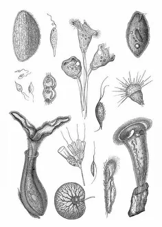
Unicellular eukaryotic organisms
Microscopic Organisms Antique Print, Vintage Lithograph, Urtiere Protozoen, Protozoa Old Zoology Illustration Unicellular Microbiology

Background imageLight Micrograph Collection: Apple stem, light micrograph

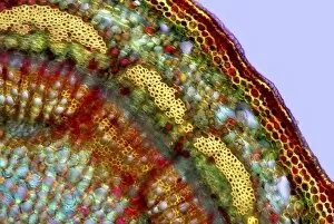
Apple stem, light micrograph
Apple stem. Polarised light micrograph of a section through the stem of a young apple (Malus domestica) tree plant. Magnification: x100 when printed 10 centimetres wide

Background imageLight Micrograph Collection: A microscope image of a desmid

A microscope image of a desmid

Background imageLight Micrograph Collection: Pituitary cells stained with eosin and haemotoxylin

Pituitary cells stained with eosin and haemotoxylin

Background imageLight Micrograph Collection: Skeletal muscle cells

Skeletal muscle cells

Background imageLight Micrograph Collection: Light microscope F006 / 7190

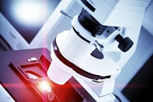
Light microscope F006 / 7190
Light microscope

Background imageLight Micrograph Collection: LM of bird flea

LM of bird flea
Light micrograph of a female flea, Ceratophylus Galinae Galinae, a parasite on dry nesting birds. The head is helmet-shaped & the body is laterally flattened

Background imageLight Micrograph Collection: LM of Demodex folliculorum

LM of Demodex folliculorum
Light micrograph in interference contrast illumination of the Follicle Mite (Demodex folliculorum). Follicle Mites are human ectoparasites, aggregating around hair follicles and in the orifices

Background imageLight Micrograph Collection: LM of HPLC column packing

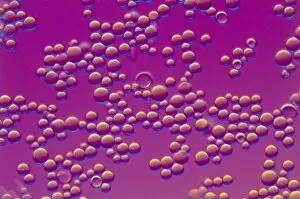
LM of HPLC column packing
Light micrograph of resin beads used in High Pressure Liquid Chromatography (HPLC), a technique for separating closely-related components from a sample solution

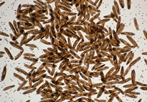
Background imageLight Micrograph Collection: LM of the protozoan Paramecium sp

LM of the protozoan Paramecium sp
Light micrograph of the ciliate protozoan, Paramecium sp. They are one of the most highly- evolved of all unicellular animals, and are plentiful in fresh water ponds

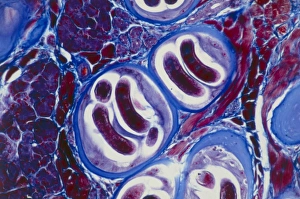
Background imageLight Micrograph Collection: LM of Trichina larvae encysted in muscle tissue

LM of Trichina larvae encysted in muscle tissue
Light micrograph of Trichina spiralis (formerly called Trichinella spiralis) larvae encysted in human muscle tissue. Each cyst (blue)

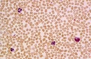
Background imageLight Micrograph Collection: LM of a field of red and white blood cells

LM of a field of red and white blood cells
Light micrograph of a sample of normal human blood, showing numerous erythrocytes (red blood cells) and four leucocytes (white blood cells) with purple-stained, lobed nuclei

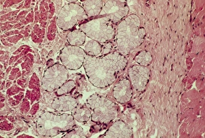
Background imageLight Micrograph Collection: LM of mammary gland

LM of mammary gland
Light micrograph of normal female breast (mammary gland) tissue from a non-pregnant woman, showing aggregations of glandular lobules

Background imageLight Micrograph Collection: Human skin section, light micrograph

Human skin section, light micrograph
Human skin. Light micrograph of a section through healthy human skin. The outer surface of the skin is at top. The uppermost surface is the epidermis

Background imageLight Micrograph Collection: LM of of a single Paramecium sp

LM of of a single Paramecium sp
Light micrograph of the ciliate protozoan Paramecium sp. The internal structure of the organism is particularly clear. The large dark area toward the top of the frame is the meganucleus

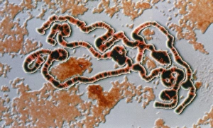
Background imageLight Micrograph Collection: LM of Drosophila giant (polytene) chromosome

LM of Drosophila giant (polytene) chromosome
Giant chromosome. Coloured computer-enhanced light micrograph of a giant (polytene) chromosome in a salivary gland cell of the fruit fly Drosophila funebris


All Professionally Made to Order for Quick Shipping

Capturing the intricate beauty of biological structures, a light micrograph reveals the mesmerizing complexity of cerebellum tissue. Delicate nerve and glial cells intertwine like a finely woven tapestry, forming the foundation for neurological function. In another stunning image, copper and magnesium sulphate crystals shimmer under the lens of a light microscope, showcasing their crystalline elegance. Moving beyond human anatomy, an awe-inspiring glimpse into early development is unveiled through a light micrograph of a human blastocyst. This embryonic stage brims with promise and potential as it prepares to embark on its journey towards life. Immunofluorescent LM unveils vibrant hues that illuminate neurons and astrocytes in breathtaking detail. These vital components of our nervous system come alive under fluorescent markers, revealing their interconnectedness in supporting brain function. Stepping away from biology but not lacking in fascination, caffeine crystals take center stage in yet another captivating light micrograph. Their jagged edges and distinct patterns mirror the stimulating effects they have on our bodies. Venturing deeper into brain tissue exploration, hippocampus tissue emerges as an enchanting subject for study. Its convoluted structure houses memories and emotions while providing insight into cognitive processes that shape who we are. Glial cells take on an ethereal quality when observed through confocal light microscopy. The interplay between these supportive cells becomes apparent as they weave together like delicate threads within neural networks. HeLa cells become protagonists under the gaze of a light microscope C017/8299 - immortalized cell lines that have revolutionized medical research since their discovery over half a century ago. Their unique characteristics continue to unlock mysteries about cancer and other diseases plaguing humanity. Intricacy extends beyond living organisms; even plant stems hold secrets waiting to be revealed by science's lens. A dicotyledon stem showcases its vascular bundles with precision while hinting at nature's ingenious design principles.