
Large Intestine Collection
The large intestine, a vital component of the human digestive system, plays a crucial role in processing waste and absorbing water from digested food


All Professionally Made to Order for Quick Shipping
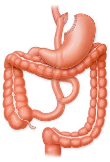
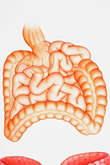
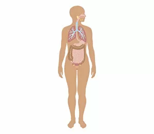
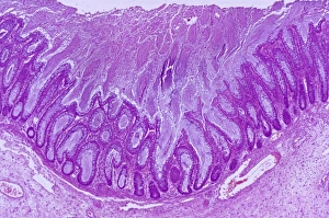
The large intestine, a vital component of the human digestive system, plays a crucial role in processing waste and absorbing water from digested food. This intricate organ can be visualized through various medical illustrations and X-rays. One such illustration showcases an X-ray of the appendix, highlighting its location within the abdomen. Another image depicts a cross-section biomedical illustration of a newborn baby boy's rib cage and intestines, providing insight into the development of this essential organ. In addition to these visuals, there is an engraving illustrating the normal situation of internal organs in both men and women. This artwork offers valuable insights into how the large intestine fits within our body structure. To further understand its function, we have an illustration depicting the entire digestive system along with common causes of diarrhea. This comprehensive diagram helps us comprehend how different parts work together to maintain overall health. Moreover, another artwork portrays healthy large intestines specifically (artwork F006 / 3476). It emphasizes that maintaining good intestinal health is crucial for optimal digestion and overall well-being. Lastly, female anatomy artwork (F008 / 1871) highlights gender-specific aspects related to this organ while emphasizing its importance in both men and women. Similarly, other artworks (F008 / 1187 & F008 / 1185) provide detailed depictions of the human digestive system as a whole. These captivating visuals not only educate but also emphasize why understanding our large intestine is critical for maintaining proper digestion and overall health.