
Lacuna Collection
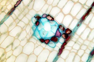
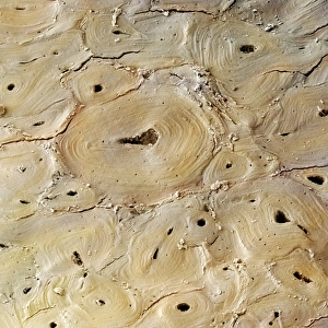
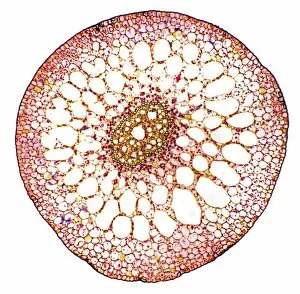
"Lacuna: Unveiling the Intricacies of Nature's Canvas" Delicate and intricate, the pine stem reveals its hidden secrets under the lens of a light micrograph


All Professionally Made to Order for Quick Shipping
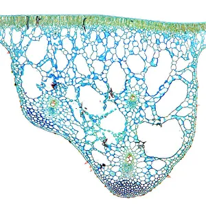
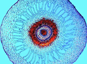
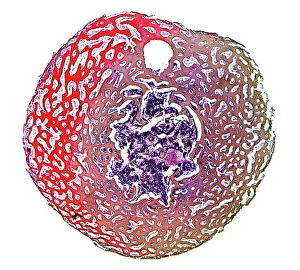
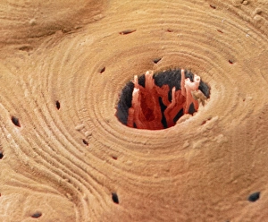
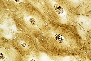
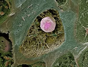
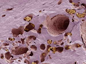
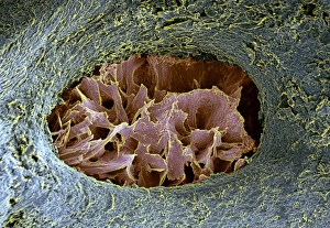
"Lacuna: Unveiling the Intricacies of Nature's Canvas" Delicate and intricate, the pine stem reveals its hidden secrets under the lens of a light micrograph. Dive into the depths of bone structure as compact bone showcases its mesmerizing patterns in a captivating light micrograph. Like a vibrant masterpiece, the water lily leaf dances with grace when examined through a light micrograph, showcasing nature's artistic touch. The water fern rhizome unravels its mysteries under the watchful eye of a light micrograph, revealing an enchanting world beneath the surface. Spongy bone comes to life in an extraordinary display captured by a light micrograph, reminding us that beauty lies even within our own bodies. Explore the elegant intricacies of pondweed stem as it unveils its hidden wonders through the lens of a mesmerizing light micrograph. "Episodes from the Life of St. Augustine" fresco takes us on an artistic journey where they are filled with stories and emotions from centuries past (1463-65). Coloured SEM offers us an unparalleled view into transverse sections of compact bone, unveiling stunning details that lie beneath our very skin. Witnessing yet another marvel through coloured SEM - this time exploring transverse sections of compact bone - reminds us how nature never ceases to amaze. A glimpse into normal human compact bone is like peering into nature's architectural masterpiece; each cell meticulously arranged for strength and resilience. In these images and artworks lies more than meets the eye – they invite us to embrace curiosity and appreciate both nature's grandeur and humanity's creative endeavors while unraveling lacunas that hold untold stories waiting to be discovered.