
Jaw Collection
"Unveiling the Marvels of the Jaw: From Leonardo da Vinci's Intricate Skull Anatomy to Ancient Fossils and Modern X-rays" Delving into the depths of anatomical knowledge


All Professionally Made to Order for Quick Shipping
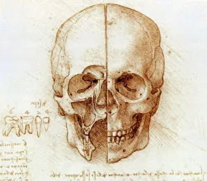
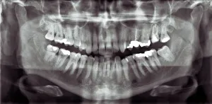
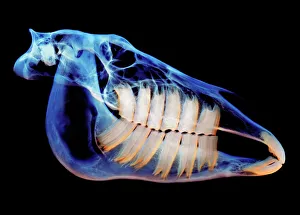
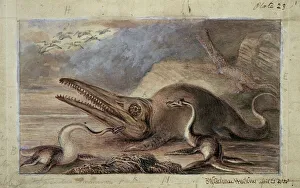
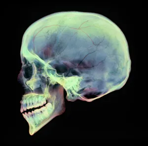
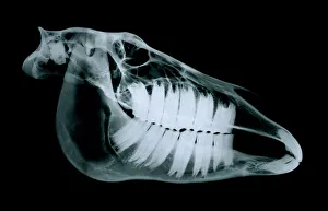
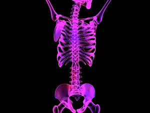
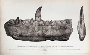
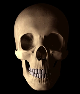
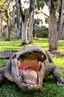
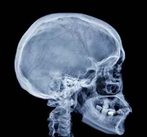
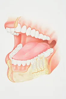
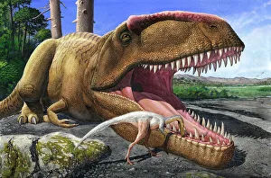
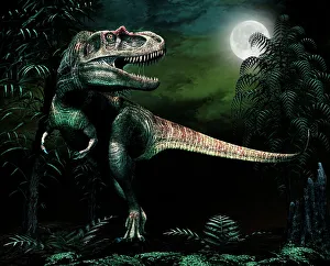
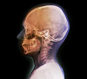
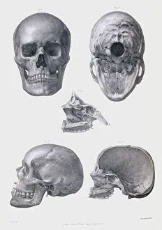
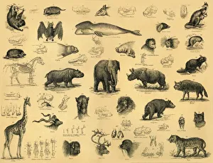
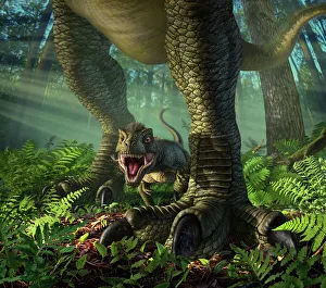
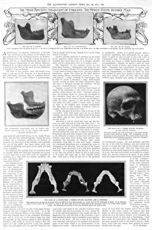
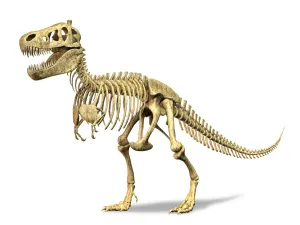
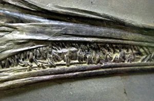
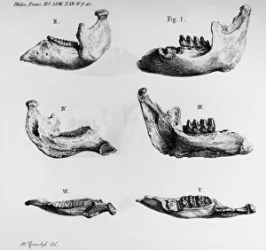
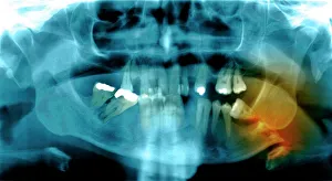
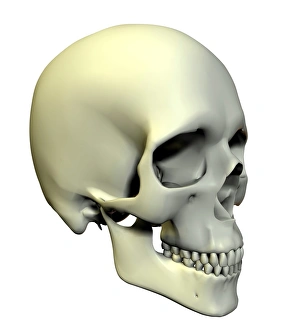
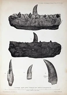
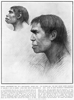
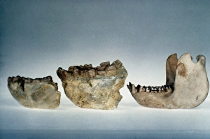
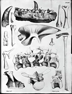
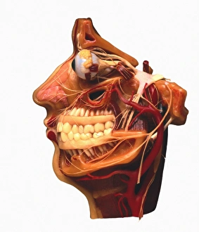
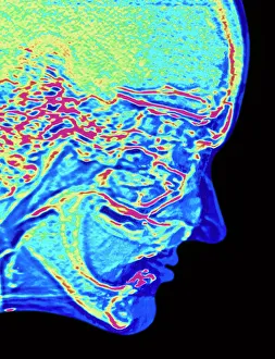
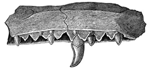
"Unveiling the Marvels of the Jaw: From Leonardo da Vinci's Intricate Skull Anatomy to Ancient Fossils and Modern X-rays" Delving into the depths of anatomical knowledge, Leonardo da Vinci's detailed sketches reveal the intricate structure of the human jaw in his renowned "Skull anatomy. " Exploring beyond our species, even horses possess a fascinating skull with a powerful jaw that enables them to graze on tough vegetation. Journeying back millions of years, fossils like Ichthyosaurus and Plesiosaurus provide glimpses into prehistoric marine reptiles' jaws, adapted for hunting and devouring prey. Advancements in technology allow us to peer inside mouths like never before - panoramic dental X-rays offer comprehensive views of teeth alignment and jaw health. Peering through an X-ray lens, we witness how human skulls showcase unique variations in jaw structure that contribute to diverse facial features. Examining equine evolution further reveals intriguing adaptations within horse skulls, highlighting their robust jaws designed for grazing and chewing grasses. Bucklands Megalosaurus jaw from 1824 provides valuable insights into early dinosaur anatomy, showcasing its formidable bite force through well-preserved fossil remains. Merging artistry with science, computer-generated imagery brings upper body skeletons to life as captivating artwork showcases intricate details of our skeletal system including the mighty jawbone. Uncovering nature's diversity yet again, American alligator C322 displays its fearsome set of jaws capable of delivering crushing bites while maintaining impressive strength throughout their lifetime. In a remarkable scene from ancient times captured by Alvarezsaurid bird cleaning Giganotosaurus carolinii's mouth; it highlights symbiotic relationships where one creature assists another by ensuring oral hygiene within massive dinosaur jaws. Cross-section diagrams illuminate the complex inner workings of our mouths and jaws – revealing how muscles align, teeth fit together, and the jawbone supports our daily activities.