Fraud Blocker Skip to main content

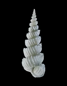
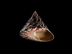
Invertebrates Collection (page 100)

"Invertebrates

Background imageInvertebrates Collection: Foraminiferan fossil, SEM

Foraminiferan fossil, SEM
Foraminiferan fossil. Coloured scanning electron micrograph (SEM) of the fossilised shell (test) of a foraminiferan. Foraminifera are single-celled marine protozoa that construct

Background imageInvertebrates Collection: LM of the protozoan Paramecium sp

LM of the protozoan Paramecium sp
Light micrograph of the ciliate protozoan, Paramecium sp. They are one of the most highly- evolved of all unicellular animals, and are plentiful in fresh water ponds

Background imageInvertebrates Collection: LM of Trichina larvae encysted in muscle tissue

LM of Trichina larvae encysted in muscle tissue
Light micrograph of Trichina spiralis (formerly called Trichinella spiralis) larvae encysted in human muscle tissue. Each cyst (blue)

Background imageInvertebrates Collection: Coloured SEM of foraminiferan, Peneroplis sp

Coloured SEM of foraminiferan, Peneroplis sp
Foraminiferan shell. Coloured scanning electron micrograph (SEM) of the test (shell) of the Mediterranean foraminiferan, Peneroplis proteus

Background imageInvertebrates Collection: Head louse claw

Head louse claw
Head louse. Coloured scanning electron micrograph (SEM) of a claw of a human head louse (Pediculus humanus capitis) clinging to a human hair

Background imageInvertebrates Collection: Salmon louse, SEM

Salmon louse, SEM
Salmon louse. Coloured scanning electron micrograph (SEM) of the underside of a salmon louse (Lepeophtheirus salmonis). This marine copepod crustacean is parasitic on salmon fish

Background imageInvertebrates Collection: SEM of Paramecium

SEM of Paramecium
Paramecium. Coloured scanning electron micrograph (SEM) of the single-celled protozoan, Paramecium caudatum. This ciliate animal has cilia on its cell membrane surface enabling it to swim with an

Background imageInvertebrates Collection: Coloured SEM of a human head louse on hair shafts

Coloured SEM of a human head louse on hair shafts
Head louse. Coloured scanning electron micrograph (SEM) of a human head louse (Pediculus humanus capitis) clinging to human hair

Background imageInvertebrates Collection: Chilodonella ciliate protozoan, SEM

Chilodonella ciliate protozoan, SEM
Chilodonella ciliate protozoa, coloured scanning electron micrograph (SEM). These tiny single- celled organisms are found in fresh water

Background imageInvertebrates Collection: LM of of a single Paramecium sp

LM of of a single Paramecium sp
Light micrograph of the ciliate protozoan Paramecium sp. The internal structure of the organism is particularly clear. The large dark area toward the top of the frame is the meganucleus

Background imageInvertebrates Collection: Coloured SEM of Podocyrtis mitra, a radiolarian

Coloured SEM of Podocyrtis mitra, a radiolarian
Radiolarian. Coloured scanning electron micrograph (SEM) of the test (skeleton) of a Podocyrtis mitra, a type of radiolarian protozoan

Background imageInvertebrates Collection: Coloured SEM of head of an earthworm

Coloured SEM of head of an earthworm
Common earthworm. Coloured scanning electron micrograph (SEM) of the head of a common earthworm (Lumbricus terrestris). At the tip of the head, at upper centre, is the mouth

Background imageInvertebrates Collection: Coloured SEM of a natural sponge

Coloured SEM of a natural sponge
Sponge. Coloured scanning electron micrograph (SEM) of an unidentified sponge, phylum Porifera. The branching structure of the sponges body is supported by an internal skeleton of calcareous or

Background imageInvertebrates Collection: Light micrograph of beef tapeworm cysts in tongue

Light micrograph of beef tapeworm cysts in tongue
Light micrograph of a section of beef tongue containing the encysted larvae of the beef tapeworm, Taenia saginata. The lower two thirds of the image (below the blue band of muscle)

Background imageInvertebrates Collection: LM of paramecium sp

LM of paramecium sp
Light micrograph in phase contrast illumination of the ciliate protozoan, Paramecium sp. The internal structure of Paramecium is visible here with many food and water vacuoles seen as bright spots

Background imageInvertebrates Collection: Coloured SEM of an earwig, Forficula auricularia

Coloured SEM of an earwig, Forficula auricularia
Earwig. Coloured scanning electron micrograph (SEM) of an earwig (Forficula auricularia) on leaf litter. Earwigs are predatory nocturnal insects which generally hide under stones

Background imageInvertebrates Collection: Barnacle glue, SEM

Barnacle glue, SEM
Barnacle glue. Coloured scanning electron micrograph (SEM) of a section through the byssus (glue threads) of an acorn barnacle (Elminius modestus)

Background imageInvertebrates Collection: Head louse

Head louse. Coloured scanning electron micrograph (SEM) of a blood-sucking human head louse (Pediculus humanus capitis) clinging to hairs with the claws on the ends of its legs

Background imageInvertebrates Collection: Protozoan sexual reproduction, SEM Z105 / 0240

Protozoan sexual reproduction, SEM Z105 / 0240
Protozoan sexual reproduction. Coloured scanning electron micrograph (SEM) of two Paramecium caudatum protozoa reproducing sexually by a method called conjugation

Background imageInvertebrates Collection: Coloured SEM of foraminiferan, Elphidium sp

Coloured SEM of foraminiferan, Elphidium sp
Foraminiferan shell. Coloured scanning electron micrograph (SEM) of the test (shell) of the British formaminiferan, Elphidium crispum

Background imageInvertebrates Collection: Coloured SEM of Foraminifera Textularia sp. fossil

Coloured SEM of Foraminifera Textularia sp. fossil
Fossil foraminifera. Coloured scanning electron micrograph (SEM) of the fossil shell of the single-celled protozoan Textularia sp. belonging to the order Foraminifera

Background imageInvertebrates Collection: Paramecium protozoa, SEM Z105 / 0239

Paramecium protozoa, SEM Z105 / 0239
Paramecium protozoa. Coloured scanning electron micrograph (SEM) of two Paramecium caudatum protozoa on filaments (green) of Oscillatoria sp. blue-green algae. P

Background imageInvertebrates Collection: Coloured SEM of a tapeworm, Taenia sp

Coloured SEM of a tapeworm, Taenia sp
Tapeworm. Coloured scanning electron micrograph (SEM) of a parasitic tapeworm (Taenia sp.). The scolex (head, at right) has suckers (upper right) and a crown of hooklets (top right)

Background imageInvertebrates Collection: Calcareous phytoplankton fossil, SEM Z100 / 0212

Calcareous phytoplankton fossil, SEM Z100 / 0212
Calcareous phytoplankton fossil. Coloured scanning electron micrograph (SEM) of a fossil of the skeleton (coccosphere) of a coccolithotrope, a small marine algal organism

Background imageInvertebrates Collection: Chilodonella ciliate protozoa, LM

Chilodonella ciliate protozoa, LM
Chilodonella protozoa. Light micrograph of several Chilodonella sp. ciliate protozoa. Chilodonella is a single-celled animal that is found in freshwater

Background imageInvertebrates Collection: Fossilised foraminiferans, SEM

Fossilised foraminiferans, SEM
Fossilised foraminiferans. Coloured scanning electron micrograph (SEM) of the fossilised shells of assorted foraminiferan protozoa (order Foraminifera)

Background imageInvertebrates Collection: Amoebae, SEM

Amoebae, SEM
Amoebae. Coloured scanning electron micrograph (SEM) of two large protozoan amoebae (Amoeba proteus). A. proteus is commonly found on the bottom of pools and puddles

Background imageInvertebrates Collection: Coloured SEM of foraminiferan, Spirolina sp

Coloured SEM of foraminiferan, Spirolina sp
Foraminiferan shell. Coloured scanning electron micrograph (SEM) of the test (shell) of the Mediterranean foraminiferan, Spirolina areitinus

Background imageInvertebrates Collection: Foraminiferan tests

Foraminiferan tests (shells), macrophotograph. Species seen here include Bulimina gibba, Elphidium crispum, E. articulatum, Lagena sulcata, and Miliolinella circularis

Background imageInvertebrates Collection: SEM of an earwig

SEM of an earwig
Earwig. Coloured scanning electron micrograph (SEM) of an earwig (Forficula auricularia) on leaf litter. Earwigs are predatory nocturnal insects which generally hide under stones

Background imageInvertebrates Collection: Acorn barnacles, SEM

Acorn barnacles, SEM
Acorn barnacles. Coloured scanning electron micrograph (SEM) of three acorn barnacles (Elminius modestus) attached to a rock

Background imageInvertebrates Collection: Foraminiferan fossil, SEM Z110 / 0239

Foraminiferan fossil, SEM Z110 / 0239
Foraminiferan fossil. Coloured scanning electron micrograph (SEM) of the fossilised shell (test) of a foraminiferan. Foraminifera are single-celled marine protozoa that construct

Background imageInvertebrates Collection: Heteropod sea snail shell C019 / 1298

Heteropod sea snail shell C019 / 1298
Heteropod sea snail shell. Shell of a Carinaria vitrea heteropod marine gastropd mollusc. The opening (left) of this shell measures 55mm in diameter

Background imageInvertebrates Collection: Volute sea snail shell C019 / 1372

Volute sea snail shell C019 / 1372
Volute sea snail shell. Shell of a volute sea snail (Volutoconus bednalli). This species of marine gastropod mollusc is in the Indo-Pacific. This specimen measures 107mm long

Background imageInvertebrates Collection: Imperial cone shell C019 / 1314

Imperial cone shell C019 / 1314
General cone shell. Shell of a general cone shell (Conus imperialis) sea snail. Cone shells are venomous predatory marine gastropod molluscs that can be dangerous to humans

Background imageInvertebrates Collection: Zigzag venus clam shell C019 / 1338

Zigzag venus clam shell C019 / 1338
Zigzag venus clam shell. Shell of a zigzag venus clam (Lioconcha castrensis). Venus clams (family Veneridae) are a very large family of minute, to large, saltwater clams and marine bivalve molluscs

Background imageInvertebrates Collection: Cockle shell C019 / 1317

Cockle shell C019 / 1317
Cockle shell. Shell of a cockle (Freneixicardia victor). This specimen was found on Mactan island, Philippines, and measure 47mm long

Background imageInvertebrates Collection: Conch shell C019 / 1337

Conch shell C019 / 1337
Conch shell. Underside of the shell of a conch (Lambis cristinae) sea snail. This species of marine gastropod mollusc is found in the Indian Ocean. This specimen measures 164mm long

Background imageInvertebrates Collection: Black-ribbed false limpet shells C019 / 1307

Black-ribbed false limpet shells C019 / 1307
Black-ribbed false limpet shells. Collection of shells from black-ribbed false limpets (Clypidina notata), showing their interior patterns

Background imageInvertebrates Collection: Slit snail shell C019 / 1294

Slit snail shell C019 / 1294
Slit snail shell. Shell of a slit snail (Bayerotrochus boucheti) sea snail. This marine gastropod mollusc is found off Norfolk Ridge, New Caledonia, in the southwest Pacific Ocean

Background imageInvertebrates Collection: Precious wentletrap sea snail shell C019 / 1324

Precious wentletrap sea snail shell C019 / 1324
Precious wentletrap sea snail shell. Shell of a precious wentletrap (Epitonium scalare) sea snail. This species of predatory marine gastropod mollusc is found in the Red Sea

Background imageInvertebrates Collection: Argonaut octopus eggcase shell C019 / 1291

Argonaut octopus eggcase shell C019 / 1291
Argonaut octopus eggcase shell. Egg case from a female greater argonaut (Argonauta argo) found in the Indo-Pacific. Argonauts are a group of pelagic octopuses

Background imageInvertebrates Collection: Harpoon carved from antler

Harpoon carved from antler. Cast of a prehistoric antler harpoon produced by the Magdalenian culture of Western Europe. This artefact, which dates from 11, 000 to 18

Background imageInvertebrates Collection: Land snail shell C019 / 1359

Land snail shell C019 / 1359
Land snail shell. Shell of a Tortulosa tortulosa land snail. This specimen measures 24mm long

Background imageInvertebrates Collection: Wentletrap sea snail shell C019 / 1325

Wentletrap sea snail shell C019 / 1325
Wentletrap sea snail shell. Shell of a precious wentletrap (Cirsostrema rugosum) sea snail. This species of predatory marine gastropod mollusc is found in the Indo-Pacific

Background imageInvertebrates Collection: Helicostoa sinensis aquatic snails C019 / 1331

Helicostoa sinensis aquatic snails C019 / 1331
Helicostoa sinensis aquatic snails. Shells from Helicostoa sinensis aquatic snails on a rock. H. sinensis are freshwater snails and the only species in the family Helicostoidae

Background imageInvertebrates Collection: Turrid sea snail shell C019 / 1368

Turrid sea snail shell C019 / 1368
Turrid sea snail shell. Shell of a turrid (Turris normandavidsoni) sea snail. This species of marine gastropod mollusc is found in the Indo-Pacific. This specimen measures 70mm long

Background imageInvertebrates Collection: Mammillaria dolichocentra cactus, artwork

Mammillaria dolichocentra cactus, artwork
William Cowper (1731-1800). English poet. Portrait. Image taken from The Rural Walks of Cowper; displayed in a series of views near Olney


All Professionally Made to Order for Quick Shipping

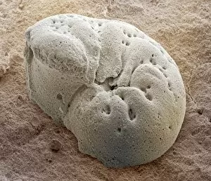
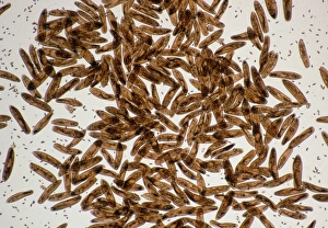
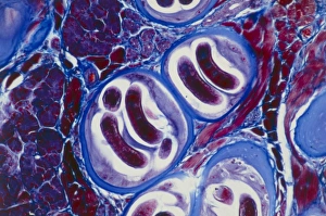
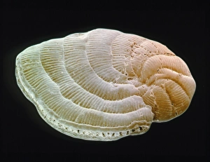
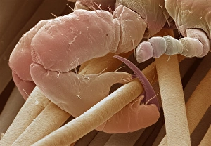
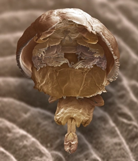
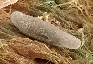
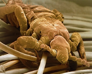
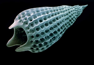
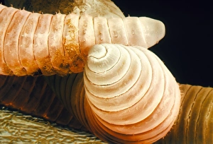
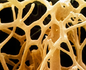
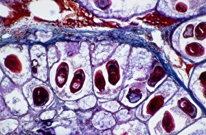
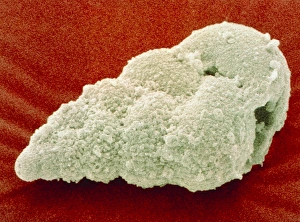
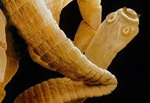
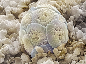
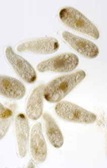
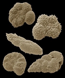
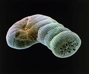
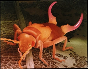
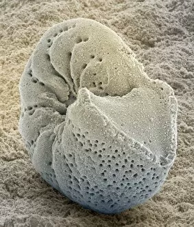
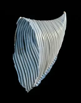
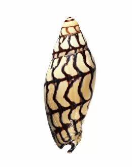
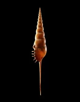
"Invertebrates: A Fascinating World of Diversity and Adaptation" From the intricate honeycombs built by diligent honey bees to the captivating sight of a lobster's inner workings revealed through an X-ray, invertebrates never cease to amaze us. The Red Admiral butterfly gracefully displays its vibrant wings, showcasing nature's artistic brilliance. Meanwhile, under the lens of a scanning electron microscope, calcareous phytoplankton reveal their delicate beauty as they float effortlessly in the vast ocean. In Kagoshima Prefecture, Kyushu, Japan, a male Leach's sea star steals the spotlight as it engages in broadcast spawning - an awe-inspiring phenomenon where streams of sperm are released from its arms into the water. This mesmerizing moment captured by a talented wildlife photographer earned them recognition as the winner in the Underwater category at Wildlife Photographer of the Year 2022. Venturing into Croatian waters along Korcula Island in the Adriatic Sea reveals another enchanting creature - a Nudibranch known as Janolus cristatus. Its vibrant colors and graceful movements make it a true gem beneath the waves. A sea green swallowtail butterfly flutters elegantly across meadows and gardens while fruit flies unveil their intricate structures when observed under high-resolution SEM imaging techniques. Delving deep into Earth's history brings us face-to-face with ancient wonders like trilobite fossils that offer glimpses into prehistoric times. These extinct arthropods remind us of our planet's rich evolutionary past. Witnessing an ocellate octopus swimming up from the depths is like witnessing magic unfold before our eyes. Its ethereal appearance leaves us captivated by nature's ability to create such extraordinary creatures. Lastly, we encounter a painted lady butterfly whose delicate patterns adorn fields with splashes of color and gracefulness. And even within microscopic realms lies astonishing complexity; for instance, observing dog tapeworm heads through SEM reveals the intricate structures that allow these parasites to thrive.