Fraud Blocker Skip to main content

Intervertebral Collection

"Exploring the Intricacies of Intervertebral: Unveiling the Backbone's Secrets" Pinned vertebrae

Background imageIntervertebral Collection: Artificial spinal disc, X-ray C016 / 6561

Artificial spinal disc, X-ray C016 / 6561
Artificial spinal disc. Coloured X-ray of the spine of a 42 year old woman, showing a prosthetic spinal disc (white) in the lumbar (lower) spine

Background imageIntervertebral Collection: Artificial spinal disc, X-ray C016 / 6562

Artificial spinal disc, X-ray C016 / 6562
Artificial spinal disc. Coloured X-ray of the spine of a 42 year old woman, showing a prosthetic spinal disc (white) in the lumbar (lower) spine

Background imageIntervertebral Collection: Computer artwork of three views of a human spine

Computer artwork of three views of a human spine
Spine. Computer illustration of three views of a healthy human spine. It is seen from the back at left, the side at centre, and from the front at right

Background imageIntervertebral Collection: Model of three vertebrae and intervertebral discs

Model of three vertebrae and intervertebral discs
Vertebrae. Model of three vertebrae from the human spine. The vertebrae are separated by intervertebral discs (grey). Vertebrae are bones with a wide

Background imageIntervertebral Collection: Human spine, three views

Human spine, three views
Spine. Computer illustration of three views of a healthy human spine. It is seen from the back at left, the side at centre, and from the front at right

Background imageIntervertebral Collection: Computer artwork of the five lumbar vertebrae

Computer artwork of the five lumbar vertebrae
Lumbar spine. Computer illustration of the five lumbar vertebrae (blue) of the human spine. The lumbar vertebrae are the largest of the unfused vertebrae in the backbone

Background imageIntervertebral Collection: Spondylitis, X-ray

Spondylitis, X-ray
Spondylitis. Coloured X-ray of the lower spine and pelvis of a patient with spondylitis, an inflammation of the synovial joints of the backbone

Background imageIntervertebral Collection: Pinned curved spine, X-ray

Pinned curved spine, X-ray
Pinned curved spine. Coloured X-ray of bones of the spine and pelvis of a 49 year old woman showing two screws (blue) pinning the lower lumbar vertebra (L5) to the sacrum of the pelvis

Background imageIntervertebral Collection: Pinned vertebrae, X-ray

Pinned vertebrae, X-ray
Pinned vertebrae. Coloured X-ray of pins (beige) joining two of the vertebrae (pink) of a 29 year old woman together to immobilise them. Vertebrae are the box-like bones that make up the spine

Background imageIntervertebral Collection: Slipped disc, CT scan

Slipped disc, CT scan
Slipped disc. Coloured computed tomography (CT) scan of an axial section through an inter- vertebral disc (orange) showing herniation

Background imageIntervertebral Collection: Ankylosing spondylitis X-ray

Ankylosing spondylitis X-ray
Ankylosing spondylitis. Coloured X-ray of the spine of a patient with ankylosing spondylitis, a form of arthritis. This is the lumbar region in the lower back

Background imageIntervertebral Collection: Slipped disc, MRI scan

Slipped disc, MRI scan
Slipped disc, coloured magnetic resonance imaging (MRI) scan. Profile view of the spine of a 59-year-old patient with severe rheumatoid arthritis and a herniated (slipped) disc (red)

Background imageIntervertebral Collection: Slipped disc, artwork

Slipped disc, artwork
Slipped disc. Artwork showing a slipped disc (orange) in a womans back. This condition is where one of the intervertebral discs (grey) in the backbone ruptures

Background imageIntervertebral Collection: Normal and slipped disc, artwork

Normal and slipped disc, artwork
Normal and slipped disc. Artwork showing a normal (top) and slipped (bottom) disc in the backbone, seen from above. The rear of the backbone (with protruding processes)

Background imageIntervertebral Collection: Intervertebral disc, light micrograph

Intervertebral disc, light micrograph
Intervertebral disc. Light micrograph of a transverse section through an intervertebral disc (red) and the surrounding tissue (brown)


All Professionally Made to Order for Quick Shipping

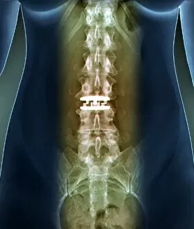
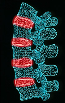
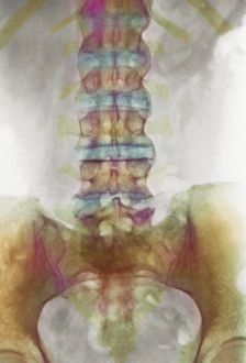
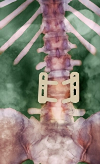
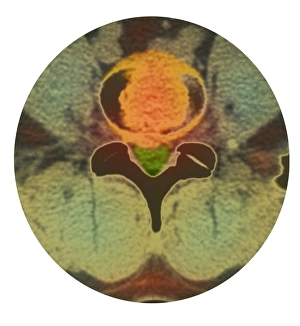
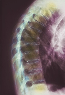
"Exploring the Intricacies of Intervertebral: Unveiling the Backbone's Secrets" Pinned vertebrae, X-ray: A close-up view connections reveals the remarkable complexity and strength that supports our spine. Artificial spinal disc, X-ray C016 / 6561: Advancements in medical technology bring hope for those suffering from spinal conditions as artificial discs provide a new lease on life. X-ray C016 / 6562: Witnessing the marvels of modern science through an x-ray image, showcasing how artificial discs seamlessly integrate into our skeletal structure. Computer artwork of three views of a human spine: Through digital artistry, we gain a comprehensive understanding of the intricate network formed by intervertebral discs within our backbone. Model of three vertebrae and intervertebral discs: A tangible representation allows us to grasp the significance and function of these crucial components that enable flexibility and movement in our spine. Human spine, three views: From various angles, we appreciate the elegance and resilience exhibited by each intervertebral connection within our complex vertebral column. Computer artwork of the five lumbar vertebrae: Delving deeper into anatomy, this visual masterpiece highlights how intervertebral structures play a pivotal role in maintaining stability while accommodating motion in our lower back region. Spondylitis, X-ray: The impact of spondylitis is unveiled through an x-ray image – reminding us to cherish healthy spines while seeking effective treatments for those affected by this condition. Pinned curved spine, X-ray (x3): Multiple perspectives capture both curiosity and concern as we observe pinned curved spines; emphasizing why early detection is vital for addressing potential issues related to intervertebral health. Intriguingly intricate yet resiliently robust.