Fraud Blocker Skip to main content

Internal Anatomy Collection (page 9)

"Exploring the Intricacies: Unveiling Internal Anatomy through Art and Science" Delving into the hidden world of internal anatomy

Background imageInternal Anatomy Collection: BARTHOLIN, Thomas (1616-1680). Danish physician, mathematician, and theologian

BARTHOLIN, Thomas (1616-1680). Danish physician, mathematician, and theologian
CUL4875648 BARTHOLIN, Thomas (1616-1680). Danish physician, mathematician, and theologian. He is best known for his work in the discovery of the lymphatic system in humans

Background imageInternal Anatomy Collection: study of a body, hand, copperplate engraving, about 1700

study of a body, hand, copperplate engraving, about 1700
UTD4921972 study of a body, hand, copperplate engraving, about 1700; (add.info.: study of a body, hand, copperplate engraving)

Background imageInternal Anatomy Collection: The eye, copperplate engraving, about 1700

The eye, copperplate engraving, about 1700
UTD4921822 The eye, copperplate engraving, about 1700; (add.info.: The eye, copperplate engraving, about 1700 ©HistAuge/United Archives/Leemage); United Archives/HistAuge

Background imageInternal Anatomy Collection: Anatomy of the human arterial system in the legs. Handcoloured lithograph by St

Anatomy of the human arterial system in the legs. Handcoloured lithograph by St
FLO4684812 Anatomy of the human arterial system in the legs. Handcoloured lithograph by St. Schillinger from Lorenz Oken's Universal Natural History, Allgemeine Naturgeschichte fur alle Stande

Background imageInternal Anatomy Collection: Anatomy of human intestines from the side. Lithograph from Lorenz Oken's Universal Natural History

Anatomy of human intestines from the side. Lithograph from Lorenz Oken's Universal Natural History
FLO4684807 Anatomy of human intestines from the side. Lithograph from Lorenz Oken's Universal Natural History, Allgemeine Naturgeschichte fur alle Stande, Stuttgart

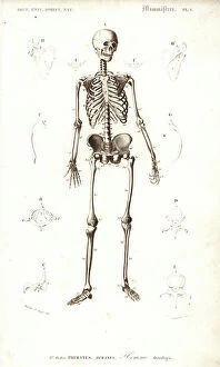
Background imageInternal Anatomy Collection: Human skeleton from the front. Engraving by Fournier after an illustration by Martin St

Human skeleton from the front. Engraving by Fournier after an illustration by Martin St
FLO4685437 Human skeleton from the front. Engraving by Fournier after an illustration by Martin St. Ange from Charles d'Orbigny's Dictionary of Natural History, Paris

Background imageInternal Anatomy Collection: Brain sections. Lithograph by Davesne, based on a drawing by Leveille

Brain sections. Lithograph by Davesne, based on a drawing by Leveille, in Petity Atlas complet d'Anatomie
FLO4673204 Brain sections. Lithograph by Davesne, based on a drawing by Leveille, in Petity Atlas complet d'Anatomie descriptive du Corps Humain, by Dr

Background imageInternal Anatomy Collection: Vertebral column, ribs, vertebrae and sternum. Lithograph of Corbie

Vertebral column, ribs, vertebrae and sternum. Lithograph of Corbie, in Petity Atlas complet d'Anatomie descriptive
FLO4673029 Vertebral column, ribs, vertebrae and sternum. Lithograph of Corbie, in Petity Atlas complet d'Anatomie descriptive du Corps Humain, by Dr

Background imageInternal Anatomy Collection: Section through the abdomen. Lithograph of Lebrun, based on a drawing by Leveille

Section through the abdomen. Lithograph of Lebrun, based on a drawing by Leveille
FLO4672840 Section through the abdomen. Lithograph of Lebrun, based on a drawing by Leveille, in Petity Atlas complet d'Anatomie descriptive du Corps Humain, by Dr

Background imageInternal Anatomy Collection: Aponevrosis (fibrous membrane that covers and supports the muscles) of the torso

Aponevrosis (fibrous membrane that covers and supports the muscles) of the torso, cross section and frontal view
FLO4672748 Aponevrosis (fibrous membrane that covers and supports the muscles) of the torso, cross section and frontal view

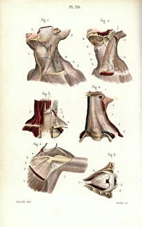
Background imageInternal Anatomy Collection: Aponevrosis of the neck, shoulder and eye. Lithograph of Corbie, based on a drawing by Leveille

Aponevrosis of the neck, shoulder and eye. Lithograph of Corbie, based on a drawing by Leveille
FLO4672728 Aponevrosis of the neck, shoulder and eye. Lithograph of Corbie, based on a drawing by Leveille, in Petiure Atlas complet d'Anatomie descriptive du Corps Humain, by Dr

Background imageInternal Anatomy Collection: Muscles and tendons of the leg and foot. Lithograph of Lebrun, based on a drawing by Leveille

Muscles and tendons of the leg and foot. Lithograph of Lebrun, based on a drawing by Leveille
FLO4672719 Muscles and tendons of the leg and foot. Lithograph of Lebrun, based on a drawing by Leveille, in Petity Atlas complet d'Anatomie descriptive du Corps Humain, by Dr

Background imageInternal Anatomy Collection: Thigh and knee muscles and tendons. Lithograph of Lebrun, based on a drawing by Leveille

Thigh and knee muscles and tendons. Lithograph of Lebrun, based on a drawing by Leveille
FLO4672703 Thigh and knee muscles and tendons. Lithograph of Lebrun, based on a drawing by Leveille, in Petity Atlas complet d'Anatomie descriptive du Corps Humain, by Dr

Background imageInternal Anatomy Collection: Thigh muscles and tendons. Lithograph of Lebrun, based on a drawing by Leveille

Thigh muscles and tendons. Lithograph of Lebrun, based on a drawing by Leveille
FLO4672702 Thigh muscles and tendons. Lithograph of Lebrun, based on a drawing by Leveille, in Petity Atlas complet d'Anatomie descriptive du Corps Humain, by Dr

Background imageInternal Anatomy Collection: Muscles and tendons of the forearm and wrist. Lithograph by Davesne, based on a drawing by Leveille

Muscles and tendons of the forearm and wrist. Lithograph by Davesne, based on a drawing by Leveille
FLO4672691 Muscles and tendons of the forearm and wrist. Lithograph by Davesne, based on a drawing by Leveille, in Petity Atlas complet d'Anatomie descriptive du Corps Humain, by Dr

Background imageInternal Anatomy Collection: Muscles of the forearm and wrist. Lithograph by Davesne, based on a drawing by Leveille

Muscles of the forearm and wrist. Lithograph by Davesne, based on a drawing by Leveille
FLO4672685 Muscles of the forearm and wrist. Lithograph by Davesne, based on a drawing by Leveille, in Petity Atlas complet d'Anatomie descriptive du Corps Humain, by Dr

Background imageInternal Anatomy Collection: Shoulder and arm muscles. Lithograph of Anneshower, based on a drawing by Leveille

Shoulder and arm muscles. Lithograph of Anneshower, based on a drawing by Leveille
FLO4672670 Shoulder and arm muscles. Lithograph of Anneshower, based on a drawing by Leveille, in Petiure Atlas complet d'Anatomie descriptive du Corps Humain, by Dr

Background imageInternal Anatomy Collection: Internal muscles of the back. Lithograph by Davesne, based on a drawing by Leveille

Internal muscles of the back. Lithograph by Davesne, based on a drawing by Leveille
FLO4672652 Internal muscles of the back. Lithograph by Davesne, based on a drawing by Leveille, in Petity Atlas complet d'Anatomie descriptive du Corps Humain, by Dr

Background imageInternal Anatomy Collection: Torso muscles (male). Lithograph of anneshower, based on a drawing by Leveille

Torso muscles (male). Lithograph of anneshower, based on a drawing by Leveille
FLO4672627 Torso muscles (male). Lithograph of anneshower, based on a drawing by Leveille, in Petiter Atlas complet d'Anatomie descriptive du Corps Humain, by Dr

Background imageInternal Anatomy Collection: Shoulder and elbow joints. Lithograph of Corbie, in Petity Atlas complet d'Anatomie descriptive du

Shoulder and elbow joints. Lithograph of Corbie, in Petity Atlas complet d'Anatomie descriptive du Corps Humain
FLO4672545 Shoulder and elbow joints. Lithograph of Corbie, in Petity Atlas complet d'Anatomie descriptive du Corps Humain, by Dr

Background imageInternal Anatomy Collection: Hand bone: carps, metacarps and phalanges and leg: femur and shin. Lithograph of Corbie

Hand bone: carps, metacarps and phalanges and leg: femur and shin. Lithograph of Corbie
FLO4672512 Hand bone: carps, metacarps and phalanges and leg: femur and shin. Lithograph of Corbie, in Petity Atlas complet d'Anatomie descriptive du Corps Humain, by Dr

Background imageInternal Anatomy Collection: Cervical plexus. Lithograph of Corbie, based on a drawing by Leveille

Cervical plexus. Lithograph of Corbie, based on a drawing by Leveille, in Petity Atlas complet d'Anatomie
FLO4673165 Cervical plexus. Lithograph of Corbie, based on a drawing by Leveille, in Petity Atlas complet d'Anatomie descriptive du Corps Humain, by Dr

Background imageInternal Anatomy Collection: Veins of the legs and foot. Lithograph of Corbie, based on a drawing by Leveille

Veins of the legs and foot. Lithograph of Corbie, based on a drawing by Leveille
FLO4673005 Veins of the legs and foot. Lithograph of Corbie, based on a drawing by Leveille, in Petity Atlas complet d'Anatomie descriptive du Corps Humain, by Dr

Background imageInternal Anatomy Collection: Axis and vertebrae. Lithograph of Corbie, in Petity Atlas complet d'Anatomie descriptive du Corps

Axis and vertebrae. Lithograph of Corbie, in Petity Atlas complet d'Anatomie descriptive du Corps Humain, by Dr
FLO4672967 Axis and vertebrae. Lithograph of Corbie, in Petity Atlas complet d'Anatomie descriptive du Corps Humain, by Dr

Background imageInternal Anatomy Collection: Circulatory system of the intestines. Lithograph of Anneshower, based on a drawing by Leveille

Circulatory system of the intestines. Lithograph of Anneshower, based on a drawing by Leveille
FLO4672922 Circulatory system of the intestines. Lithograph of Anneshower, based on a drawing by Leveille, in Petity Atlas complet d'Anatomie descriptive du Corps Humain, by Dr

Background imageInternal Anatomy Collection: Back and torso muscles. Lithograph by Davesne, based on a drawing by Leveille

Back and torso muscles. Lithograph by Davesne, based on a drawing by Leveille
FLO4672626 Back and torso muscles. Lithograph by Davesne, based on a drawing by Leveille, in Petity Atlas complet d'Anatomie descriptive du Corps Humain, by Dr

Background imageInternal Anatomy Collection: Pelvic bone, front and back views. Lithograph of Corbie, in Petity Atlas complet d'Anatomie

Pelvic bone, front and back views. Lithograph of Corbie, in Petity Atlas complet d'Anatomie descriptive du Corps
FLO4672563 Pelvic bone, front and back views. Lithograph of Corbie, in Petity Atlas complet d'Anatomie descriptive du Corps Humain, by Dr

Background imageInternal Anatomy Collection: Vertebral column, spine and neck. Lithograph of Corbie, in Petity Atlas complet d'Anatomie

Vertebral column, spine and neck. Lithograph of Corbie, in Petity Atlas complet d'Anatomie descriptive du Corps
FLO4672546 Vertebral column, spine and neck. Lithograph of Corbie, in Petity Atlas complet d'Anatomie descriptive du Corps Humain, by Dr

Background imageInternal Anatomy Collection: Glossopharyngeal nerve. Lithograph by Davesne, based on a drawing by Leveille

Glossopharyngeal nerve. Lithograph by Davesne, based on a drawing by Leveille
FLO4672515 Glossopharyngeal nerve. Lithograph by Davesne, based on a drawing by Leveille, in Petity Atlas complet d'Anatomie descriptive du Corps Humain, by Dr

Background imageInternal Anatomy Collection: Nerves of the skull and eye. Lithograph by Davesne, based on a drawing by Leveille

Nerves of the skull and eye. Lithograph by Davesne, based on a drawing by Leveille
FLO4672497 Nerves of the skull and eye. Lithograph by Davesne, based on a drawing by Leveille, in Petity Atlas complet d'Anatomie descriptive du Corps Humain, by Dr

Background imageInternal Anatomy Collection: Ophthalmic and maxillary nerves of the fifth cranial nerve. Lithograph by Davesne

Ophthalmic and maxillary nerves of the fifth cranial nerve. Lithograph by Davesne, based on a drawing by Leveille
FLO4672496 Ophthalmic and maxillary nerves of the fifth cranial nerve. Lithograph by Davesne, based on a drawing by Leveille, in Petity Atlas complet d'Anatomie descriptive du Corps Humain

Background imageInternal Anatomy Collection: Lumbar plexus and crural nerve. Lithograph of Corbie, based on a drawing by Leveille

Lumbar plexus and crural nerve. Lithograph of Corbie, based on a drawing by Leveille
FLO4672455 Lumbar plexus and crural nerve. Lithograph of Corbie, based on a drawing by Leveille, in Petity Atlas complet d'Anatomie descriptive du Corps Humain, by Dr

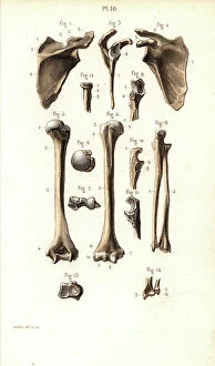
Background imageInternal Anatomy Collection: Shoulder and arm bone: scapula, humerus, cubitus and radius. Lithograph of Corbie

Shoulder and arm bone: scapula, humerus, cubitus and radius. Lithograph of Corbie
FLO4672433 Shoulder and arm bone: scapula, humerus, cubitus and radius. Lithograph of Corbie, in Petity Atlas complet d'Anatomie descriptive du Corps Humain, by Dr

Background imageInternal Anatomy Collection: Side view of human skeleton and horse skeleton. Copperplate engraving by Edward Mitchell after

Side view of human skeleton and horse skeleton. Copperplate engraving by Edward Mitchell after anatomical illustrations
FLO4565764 Side view of human skeleton and horse skeleton. Copperplate engraving by Edward Mitchell after anatomical illustrations by Bernhard Siegfried Albinus

Background imageInternal Anatomy Collection: Fetal skeleton at six months with scythe 1, at three months 2 and at ten weeks 3

Fetal skeleton at six months with scythe 1, at three months 2 and at ten weeks 3
FLO4565763 Fetal skeleton at six months with scythe 1, at three months 2 and at ten weeks 3. Copperplate engraving by Edward Mitchell after an anatomical illustration by Jean-Joseph Sue from John

Background imageInternal Anatomy Collection: Fetal skeleton at the time of birth. Copperplate engraving by Edward Mitchell after an anatomical

Fetal skeleton at the time of birth. Copperplate engraving by Edward Mitchell after an anatomical illustration by
FLO4565750 Fetal skeleton at the time of birth. Copperplate engraving by Edward Mitchell after an anatomical illustration by Jean-Joseph Sue from John Barclay's A Series of Engravings of

Background imageInternal Anatomy Collection: Asthma. Engraving from ' Les Grands Ales et les Grands Remedes' by Jules Rengade, 1889

Asthma. Engraving from " Les Grands Ales et les Grands Remedes" by Jules Rengade, 1889
PCT4274721 Asthma. Engraving from " Les Grands Ales et les Grands Remedes" by Jules Rengade, 1889.; (add.info.: Asthma)

Background imageInternal Anatomy Collection: The Canon of Medicine by Avicenna

The Canon of Medicine by Avicenna
2625213 The Canon of Medicine by Avicenna; (add.info.: The central text consists of the anatomical sections of the Canon of Medicine by Avicenna)

Background imageInternal Anatomy Collection: Equine anatomical Illustration

Equine anatomical Illustration
2626318 Equine anatomical Illustration; (add.info.: Equine anatomical Illustration from Anatomia del cavallo, infermita, et suoi rimedii')

Background imageInternal Anatomy Collection: Equine anatomical Illustration

Equine anatomical Illustration
2626316 Equine anatomical Illustration; (add.info.: Equine anatomical Illustration from Anatomia del cavallo, infermita, et suoi rimedii')

Background imageInternal Anatomy Collection: Equine anatomical Illustration

Equine anatomical Illustration
2626314 Equine anatomical Illustration; (add.info.: Equine anatomical Illustration from Anatomia del cavallo, infermita, et suoi rimedii')

Background imageInternal Anatomy Collection: Equine anatomical Illustration

Equine anatomical Illustration
2626312 Equine anatomical Illustration; (add.info.: Equine anatomical Illustration from Anatomia del cavallo, infermita, et suoi rimedii')

Background imageInternal Anatomy Collection: Shi si jing fa hui

Shi si jing fa hui
2626543 Shi si jing fa hui; (add.info.: Illustration from the Japanese & Chinese edition (1716) of Shi si jing fa hui, (Routes of the Fourteen Meridians and their Functions)

Background imageInternal Anatomy Collection: A set of anatomical tables, with explanations, and an abridgment, of the practice of midwifery

A set of anatomical tables, with explanations, and an abridgment, of the practice of midwifery, 18th (engraving)
2626224 A set of anatomical tables, with explanations, and an abridgment, of the practice of midwifery, 18th (engraving); (add.info)

Background imageInternal Anatomy Collection: A set of anatomical tables, with explanations, and an abridgment, of the practice of midwifery

A set of anatomical tables, with explanations, and an abridgment, of the practice of midwifery, 18th (engraving)
2626223 A set of anatomical tables, with explanations, and an abridgment, of the practice of midwifery, 18th (engraving); (add.info)

Background imageInternal Anatomy Collection: A set of anatomical tables, with explanations, and an abridgment, of the practice of midwifery

A set of anatomical tables, with explanations, and an abridgment, of the practice of midwifery, 18th (engraving)
2626220 A set of anatomical tables, with explanations, and an abridgment, of the practice of midwifery, 18th (engraving); (add.info)

Background imageInternal Anatomy Collection: Fasciculus medicinae

Fasciculus medicinae
2626474 Fasciculus medicinae; (add.info.: Illustration from Fasciculus medicinae Venice; 1494 the first printed book to contain anatomical illustrations)

Background imageInternal Anatomy Collection: Les oeuures d'Ambroise Pare?

Les oeuures d'Ambroise Pare?
2626406 Les oeuures d'Ambroise Pare?; (add.info.: Illustration from Les oeuures d'Ambroise Pare? 1585 by Ambroise Pare?, 1510-1590 French surgeon and anatomist.); Universal History Archive/UIG


All Professionally Made to Order for Quick Shipping

"Exploring the Intricacies: Unveiling Internal Anatomy through Art and Science" Delving into the hidden world of internal anatomy, we are captivated by the wonders that lie beneath our skin. From the delicate cross-section of a Honey Bee (Apis mellifera) to the meticulous illustrations of the Channel Tunnel Project, each piece unravels a unique perspective on this captivating subject. Intriguingly, even centuries ago, artists like Giovanni Battista Bianchi skillfully captured human form in terracotta sculptures, reminding us of our intricate inner workings. Meanwhile, Andreas Vesalius revolutionized anatomical understanding with his groundbreaking studies during the Renaissance era. As we journey through time and mediums, Waldeck's original artwork takes us back to Mesoamerica in 1831. The prints from "La nature et l'homme" by Rengade depict twin fetuses in their ideal position during gemellar pregnancy – a testament to both artistry and scientific observation. The diversity within these artworks is astounding; from portraits such as Lucrezia Panciatichi's enigmatic gaze immortalized in oil on wood panel to Giulia di Alessandro de Medici holding a book – perhaps symbolizing knowledge about one's own body. Even photography joins this exploration as two male nudes perch upon a rock in an evocative black-and-white image from 1890. This photograph captures not only physicality but also invites contemplation on vulnerability and strength intertwined within our internal structures. From ancient times to modern endeavors like the Channel Tunnel Project illustration from 1906, humanity has sought to unravel its own mysteries. Through art and science alike, we continue to marvel at our internal anatomy – an ever-evolving tapestry waiting for discovery.