Fraud Blocker Skip to main content

Internal Anatomy Collection

"Exploring the Intricacies: Unveiling Internal Anatomy through Art and Science" Delving into the hidden world of internal anatomy

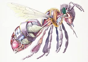
Background imageInternal Anatomy Collection: Honey Bee (Apis mellifera), internal anatomy, cross-section

Honey Bee (Apis mellifera), internal anatomy, cross-section

Background imageInternal Anatomy Collection: Karl Ernst Bock, 1809-1874 (engraving)

Karl Ernst Bock, 1809-1874 (engraving)
8632224 Karl Ernst Bock, 1809-1874 (engraving) by German School, (19th century) (after); Private Collection; (add.info.: Karl Ernst Bock, 1809-1874)

Suspensory muscle of the duodenum.Small intestine, 1905 (engraving)
8766911 Suspensory muscle of the duodenum.Small intestine, 1905 (engraving); Private Collection; (add.info.: Suspensory muscle of the duodenum.Small intestine. Human Anatomy by L)

Cross section of a melanoid cancer, 1867 (engraving)
8766908 Cross section of a melanoid cancer, 1867 (engraving); Private Collection; (add.info.: Cross section of a melanoid cancer ( Dr. Jaccoud dictionary . Vol)

Background imageInternal Anatomy Collection: Engraving of a pregnant woman's body, 1498, Johannes de Ketham

Engraving of a pregnant woman's body, 1498, Johannes de Ketham
AIS5390851 Engraving of a pregnant woman's body, 1498, Johannes de Ketham; Biblioteca Nacional, Madrid, Spain; (add.info.: Description of a pregnant woman's body)

Background imageInternal Anatomy Collection: Breast, tongue, skin and finger sections. Lithograph of Corbie, based on a drawing by Leveille

Breast, tongue, skin and finger sections. Lithograph of Corbie, based on a drawing by Leveille
FLO4672795 Breast, tongue, skin and finger sections. Lithograph of Corbie, based on a drawing by Leveille, in Petity Atlas complet d'Anatomie descriptive du Corps Humain, by Dr

Background imageInternal Anatomy Collection: Neck, tongue and throat muscles. Lithograph of Choubard, based on a drawing by Leveille

Neck, tongue and throat muscles. Lithograph of Choubard, based on a drawing by Leveille
FLO4672612 Neck, tongue and throat muscles. Lithograph of Choubard, based on a drawing by Leveille, in Petity Atlas complet d'Anatomie descriptive du Corps Humain, by Dr

Background imageInternal Anatomy Collection: Vertical sectionof the mucous membrane, 1905 (engraving)

Vertical sectionof the mucous membrane, 1905 (engraving)
8766912 Vertical sectionof the mucous membrane, 1905 (engraving); Private Collection; (add.info.: Vertical sectionof the mucous membrane, end of stomach and beginning of duodenum)

Background imageInternal Anatomy Collection: Fish-skin helmet, c.1800-1900 (fish skin)

Fish-skin helmet, c.1800-1900 (fish skin)
7264988 Fish-skin helmet, c.1800-1900 (fish skin); Manchester Museum, The University of Manchester, England; (add.info.: Helmet made from a large Porcupine fish)

Background imageInternal Anatomy Collection: Monstrum acephalon : Woodcut, Monstrum acephalon, monster without a head

Monstrum acephalon : Woodcut, Monstrum acephalon, monster without a head. From the 1642 book Monstrorum Historia by
UIG5327113 Monstrum acephalon : Woodcut, Monstrum acephalon, monster without a head. From the 1642 book Monstrorum Historia by Ulisse Aldrovandi (Bologna, 1522-1605)

Aries Biceps : Renaissance woodcut Ram. From the 1642 book Monstrorum Historia by Ulisse Aldrovandi (Bologna)
UIG5327069 Aries Biceps : Renaissance woodcut Ram. From the 1642 book Monstrorum Historia by Ulisse Aldrovandi (Bologna, 1522-1605)

Lupus Marinus Lupus Scythius : This creature looks a bit like a lion with a baboon's nose
UIG5327065 Lupus Marinus Lupus Scythius : This creature looks a bit like a lion with a baboon's nose and mane well-tamed by conditioner. "Lupus," however, suggests that it's a wolf

Harpy Secunda : Harpyae secunda icon, Egyptian harpy. Renaissance Woodcut monsters
UIG5327064 Harpy Secunda : Harpyae secunda icon, Egyptian harpy. Renaissance Woodcut monsters. From the 1642 book Monstrorum Historia by Ulisse Aldrovandi (Bologna, 1522-1605)

Background imageInternal Anatomy Collection: Puer Cornutus : Puer cornutus: a child with horns. From the 1642 book Monstrorum Historia by

Puer Cornutus : Puer cornutus: a child with horns. From the 1642 book Monstrorum Historia by Ulisse Aldrovandi
UIG5327158 Puer Cornutus : Puer cornutus: a child with horns. From the 1642 book Monstrorum Historia by Ulisse Aldrovandi (Bologna, 1522-1605)

Sus Capite & Pedibus Humanis : Sus capite & pedibus humanis, pig with human head and hands
UIG5327153 Sus Capite & Pedibus Humanis : Sus capite & pedibus humanis, pig with human head and hands. From the 1642 book Monstrorum Historia by Ulisse Aldrovandi (Bologna, 1522-1605)

Background imageInternal Anatomy Collection: Pullus Equinus Humana Facie : Horse with human face. From the 1642 book Monstrorum Historia by

Pullus Equinus Humana Facie : Horse with human face. From the 1642 book Monstrorum Historia by Ulisse Aldrovandi
UIG5327149 Pullus Equinus Humana Facie : Horse with human face. From the 1642 book Monstrorum Historia by Ulisse Aldrovandi (Bologna, 1522-1605)

Four Beasts : Hypertrichosis (also called Ambras syndrome) is an abnormal amount of hair growth over the body;
UIG5327174 Four Beasts : Hypertrichosis (also called Ambras syndrome) is an abnormal amount of hair growth over the body; extensive cases of hypertrichosis have informally been called werewolf

Agnus capite monstrifico : Agnus capite monstrifico, lamb with monstrous head
UIG5327169 Agnus capite monstrifico : Agnus capite monstrifico, lamb with monstrous head. From the 1642 book Monstrorum Historia by Ulisse Aldrovandi (Bologna, 1522-1605)

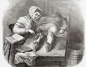
Background imageInternal Anatomy Collection: Medical College for Women, New York

Medical College for Women, New York
7301631 Medical College for Women, New York.; Private Collection; (add.info.: A student dissecting a leg at the Medical College for Women on East Twelfth Street and Second Avenue, New York, USA)

Background imageInternal Anatomy Collection: Anatomical section of the head

Anatomical section of the head
6266170 Anatomical section of the head; (add.info.: Anatomical section of the head. After a 19th century study by Robert Muys.); Photo © Ken Welsh. All rights reserved 2024.

Background imageInternal Anatomy Collection: Lo spellato' (The bark) Bronze sculpture by Ludovico Cardi called Le Cigoli (1559-1613) Florence

Lo spellato' (The bark) Bronze sculpture by Ludovico Cardi called Le Cigoli (1559-1613) Florence
BEN5479689 Lo spellato' (The bark) Bronze sculpture by Ludovico Cardi called Le Cigoli (1559-1613) Florence. Museo Nazionale del Bargello by Cardi

Background imageInternal Anatomy Collection: Lo spellato' (The bark) Bronze sculpture by Ludovico Cardi called Le Cigoli (1559-1613) Florence

Lo spellato' (The bark) Bronze sculpture by Ludovico Cardi called Le Cigoli (1559-1613) Florence
BEN5479860 Lo spellato' (The bark) Bronze sculpture by Ludovico Cardi called Le Cigoli (1559-1613) Florence. Museo Nazionale del Bargello by Cardi

Anatomy: 'knee joint' Wax sculpture by Clemente Susini (1757-1814) Museo della Specola. florence
BEN5478733 Anatomy: 'knee joint' Wax sculpture by Clemente Susini (1757-1814) Museo della Specola. florence by Susini, Clemente (1754-1814); Photo © Raffaello Bencini.

Map of nerve bundles in the human brain by Paul Broca
5309460 Map of nerve bundles in the human brain by Paul Broca; (add.info.: Map of nerve bundles in the human brain by Paul Broca)

Different views of the human heart
5308008 Different views of the human heart.; (add.info.: Print depicting different views of the human heart by Bartolomeo Eustachi (d.1574) one of the founders of the science of human anatomy

Background imageInternal Anatomy Collection: Octopus anatomy, ventral face, 1900 (engraving)

Octopus anatomy, ventral face, 1900 (engraving)
6947882 Octopus anatomy, ventral face, 1900 (engraving); Private Collection; (add.info.: Octopus anatomy , ventral face, 1900); Photo ©Jaime Abecasis.

Background imageInternal Anatomy Collection: Jaw of Lachesis muta (Grage Grand Carreau or master of the bush - venomous pit viper species jaw)

Jaw of Lachesis muta (Grage Grand Carreau or master of the bush - venomous pit viper species jaw)
CSO4996981 Jaw of Lachesis muta (Grage Grand Carreau or master of the bush - venomous pit viper species jaw), Museum d'histoire naturelle de Marseille; (add.info)

Advertisement for Ashton Bennett Magnetic Corset Belts, 1923 (litho)
8641318 Advertisement for Ashton Bennett Magnetic Corset Belts, 1923 (litho) by English School, (20th century); Private Collection; (add.info.: Advertisement for Ashton Bennett Magnetic Corset Belts)

Background imageInternal Anatomy Collection: Human skeleton (coloured engraving)

Human skeleton (coloured engraving)
8655714 Human skeleton (coloured engraving) by French School, (19th century) (after); Private Collection; (add.info.: Human skeleton)

Background imageInternal Anatomy Collection: Internal organs of a normal, healthy adult man (coloured engraving)

Internal organs of a normal, healthy adult man (coloured engraving)
8655763 Internal organs of a normal, healthy adult man (coloured engraving) by French School, (19th century) (after); Private Collection; (add.info.: Internal organs of a normal, healthy adult man)

Background imageInternal Anatomy Collection: Intestinal absorption in the digestive system (coloured engraving)

Intestinal absorption in the digestive system (coloured engraving)
8655742 Intestinal absorption in the digestive system (coloured engraving) by French School, (19th century) (after); Private Collection; (add.info.: Intestinal absorption in the digestive system)

Structure of the heart (coloured engraving)
8655735 Structure of the heart (coloured engraving) by French School, (19th century) (after); Private Collection; (add.info.: Structure of the heart)

Nerves of the face (coloured engraving)
8655732 Nerves of the face (coloured engraving) by French School, (19th century) (after); Private Collection; (add.info.: Nerves of the face)

Brain viewed in profile and cross-section (coloured engraving)
8655728 Brain viewed in profile and cross-section (coloured engraving) by French School, (19th century) (after); Private Collection; (add.info.: Brain viewed in profile and cross-section)

Background imageInternal Anatomy Collection: Connection of the sympathetic trunk with the vagus nerve and the spinal nerves (coloured engraving)

Connection of the sympathetic trunk with the vagus nerve and the spinal nerves (coloured engraving)
8655724 Connection of the sympathetic trunk with the vagus nerve and the spinal nerves (coloured engraving) by French School

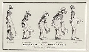
Background imageInternal Anatomy Collection: Evolution of the human skeleton (litho)

Evolution of the human skeleton (litho)
8672117 Evolution of the human skeleton (litho) by English School, (19th century); Private Collection; (add.info.: Evolution of the human skeleton, as described by Thomas Henry Huxley)

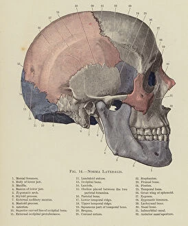
Background imageInternal Anatomy Collection: Norma Lateralis (colour litho)

Norma Lateralis (colour litho)
8667626 Norma Lateralis (colour litho) by English School, (20th century); Private Collection; (add.info.: Norma Lateralis)

Background imageInternal Anatomy Collection: Anterior View of Elbow Joint (colour litho)

Anterior View of Elbow Joint (colour litho)
8667622 Anterior View of Elbow Joint (colour litho) by English School, (20th century); Private Collection; (add.info.: Anterior View of Elbow Joint)

Background imageInternal Anatomy Collection: Dissection of the Knee Joint from the Front, Patella thrown down (colour litho)

Dissection of the Knee Joint from the Front, Patella thrown down (colour litho)
8667620 Dissection of the Knee Joint from the Front, Patella thrown down (colour litho) by English School, (20th century); Private Collection; (add.info.: Dissection of the Knee Joint from the Front)

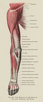
Background imageInternal Anatomy Collection: The Muscles on the Back of the Arm, Forearm, and Hand (colour litho)

The Muscles on the Back of the Arm, Forearm, and Hand (colour litho)
8667619 The Muscles on the Back of the Arm, Forearm, and Hand (colour litho) by English School, (20th century); Private Collection; (add.info.: The Muscles on the Back of the Arm, Forearm, and Hand)

Background imageInternal Anatomy Collection: Motor Points of the Upper Extremity, Anterior Aspect (litho)

Motor Points of the Upper Extremity, Anterior Aspect (litho)
8667614 Motor Points of the Upper Extremity, Anterior Aspect (litho) by English School, (20th century); Private Collection; (add.info.: Motor Points of the Upper Extremity, Anterior Aspect)

Background imageInternal Anatomy Collection: engraving of the book of Ambroise Pare: Des Monsters et Prodiges (first edition 1573)

engraving of the book of Ambroise Pare: Des Monsters et Prodiges (first edition 1573)
PCT4260992 engraving of the book of Ambroise Pare: Des Monsters et Prodiges (first edition 1573, appendix: Monsters marine) - Triton et Sirene du Nile; (add.info)

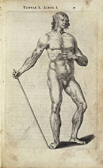
Background imageInternal Anatomy Collection: study of a body, hand, copperplate engraving, around 1700

study of a body, hand, copperplate engraving, around 1700
UTD4923684 study of a body, hand, copperplate engraving, around 1700; (add.info.: study of a body, hand, copperplate engraving)

BARTHOLIN, Thomas (1616-1680). Danish physician, mathematician, and theologian
CUL4875538 BARTHOLIN, Thomas (1616-1680). Danish physician, mathematician, and theologian. He is best known for his work in the discovery of the lymphatic system in humans

Background imageInternal Anatomy Collection: Human Anatomy book by MONTANA DE MONSERRATE, Bernardino, 1551 (engraving)

Human Anatomy book by MONTANA DE MONSERRATE, Bernardino, 1551 (engraving)
CUL4932051 Human Anatomy book by MONTANA DE MONSERRATE, Bernardino, 1551 (engraving) by Spanish School, (16th century); Biblioteca Nacional, Madrid, Spain; (add.info.: MONTANA DE MONSERRATE)

Background imageInternal Anatomy Collection: Human Anatomy book by MONTANA DE MONSERRATE, Bernardino, 1551 (engraving)

Human Anatomy book by MONTANA DE MONSERRATE, Bernardino, 1551 (engraving)
CUL4932007 Human Anatomy book by MONTANA DE MONSERRATE, Bernardino, 1551 (engraving); Biblioteca Nacional, Madrid, Spain; (add.info.: MONTANA DE MONSERRATE, Bernardino (1480-1588). Spanish doctor

Background imageInternal Anatomy Collection: Anatomical Plate - in ' De anatome corporis humani' by Guido Guidi (Vidus Vidius) (1509-1569)

Anatomical Plate - in " De anatome corporis humani" by Guido Guidi (Vidus Vidius) (1509-1569)
XEE5029671 Anatomical Plate - in " De anatome corporis humani" by Guido Guidi (Vidus Vidius) (1509-1569) Italian surgeon and anatomist

Background imageInternal Anatomy Collection: Anatomical plate in ' De anatome corporis humani' by Guido Guidi (Vidus Vidius) (1509-1569)

Anatomical plate in " De anatome corporis humani" by Guido Guidi (Vidus Vidius) (1509-1569)
XEE5029670 Anatomical plate in " De anatome corporis humani" by Guido Guidi (Vidus Vidius) (1509-1569) Italian surgeon and anatomist


All Professionally Made to Order for Quick Shipping

"Exploring the Intricacies: Unveiling Internal Anatomy through Art and Science" Delving into the hidden world of internal anatomy, we are captivated by the wonders that lie beneath our skin. From the delicate cross-section of a Honey Bee (Apis mellifera) to the meticulous illustrations of the Channel Tunnel Project, each piece unravels a unique perspective on this captivating subject. Intriguingly, even centuries ago, artists like Giovanni Battista Bianchi skillfully captured human form in terracotta sculptures, reminding us of our intricate inner workings. Meanwhile, Andreas Vesalius revolutionized anatomical understanding with his groundbreaking studies during the Renaissance era. As we journey through time and mediums, Waldeck's original artwork takes us back to Mesoamerica in 1831. The prints from "La nature et l'homme" by Rengade depict twin fetuses in their ideal position during gemellar pregnancy – a testament to both artistry and scientific observation. The diversity within these artworks is astounding; from portraits such as Lucrezia Panciatichi's enigmatic gaze immortalized in oil on wood panel to Giulia di Alessandro de Medici holding a book – perhaps symbolizing knowledge about one's own body. Even photography joins this exploration as two male nudes perch upon a rock in an evocative black-and-white image from 1890. This photograph captures not only physicality but also invites contemplation on vulnerability and strength intertwined within our internal structures. From ancient times to modern endeavors like the Channel Tunnel Project illustration from 1906, humanity has sought to unravel its own mysteries. Through art and science alike, we continue to marvel at our internal anatomy – an ever-evolving tapestry waiting for discovery.