Fraud Blocker Skip to main content

Infecting Collection

"Infecting the Microscopic World

Background imageInfecting Collection: Bacteria infecting a macrophage, SEM

Bacteria infecting a macrophage, SEM
Bacteria infecting a macrophage. Coloured scanning electron micrograph (SEM) of Mycobacterium tuberculosis bacteria (purple) infecting a macrophage white blood cell

Background imageInfecting Collection: Hepatitis C viruses, TEM

Hepatitis C viruses, TEM
Hepatitis C viruses. Coloured transmission electron micrograph (TEM) of hepatitis C virus particles (green) infecting cultured liver cells (purple)

Background imageInfecting Collection: Mouse malaria parasite, SEM

Mouse malaria parasite, SEM
Mouse malaria parasite. Coloured scanning electron micrograph (SEM) of a Plasmodium berghei protozoan (yellow) and red blood cells. P. berghei is the parasite that causes malaria in mice

Background imageInfecting Collection: Rift Valley fever virus, TEM

Rift Valley fever virus, TEM

Background imageInfecting Collection: Bacteria infecting a macrophage, SEM

Bacteria infecting a macrophage, SEM
Bacteria infecting a macrophage. Coloured scanning electron micrograph (SEM) of Mycobacterium tuberculosis bacteria (purple) infecting a macrophage white blood cell

Background imageInfecting Collection: The crimes of ignorance, the people of Molfetta destroy a cart of disinfectants intended for

The crimes of ignorance, the people of Molfetta destroy a cart of disinfectants intended for... (colour litho)
2815377 The crimes of ignorance, the people of Molfetta destroy a cart of disinfectants intended for... (colour litho) by Beltrame

Background imageInfecting Collection: Cerberus, chained by Hercules, infects the Earth with his foam, 1774. Artist: W Walker

Cerberus, chained by Hercules, infects the Earth with his foam, 1774. Artist: W Walker
Cerberus, chained by Hercules, infects the Earth with his foam, 1774. A plate from The Copper-Plate Magazine or A Monthly Treasure, London, 1774

Background imageInfecting Collection: Rust fungus infection, SEM

Rust fungus infection, SEM
Rust fungus infection. Coloured scanning electron micrograph (SEM) of spore-bearing aecia from a rust (Puccinia lagenophora) fungus, on a common groundsel (Senecio vulgaris) plant

Background imageInfecting Collection: HIV infected macrophage, SEM C018 / 8598

HIV infected macrophage, SEM C018 / 8598
HIV infected macrophage. Coloured ion-abrasion scanning electron micrograph (IA-SEM) of a macrophage white blood cell infected with human immunodeficiency virus (HIV, red)

Background imageInfecting Collection: HIV viruses, TEM

HIV viruses, TEM
HIV viruses. Coloured transmission electron micrograph (TEM) of human immunodeficiency virus (HIV) particles. These particles are docking at a T-lymphocyte (white blood cell) just before infection

Background imageInfecting Collection: Bacterial lung infection

Bacterial lung infection, computer artwork. Rod-shaped bacteria (bacilli, green) on the surface of the alveoli of the lungs (purple)

Background imageInfecting Collection: Common Ash (Fraxinus excelsior) dieback of leaves, caused by Ash Dieback (Chalara fraxinea)

Common Ash (Fraxinus excelsior) dieback of leaves, caused by Ash Dieback (Chalara fraxinea) fungal disease, Norfolk, England, June

Background imageInfecting Collection: Chalara dieback of ash sign in woodland with trees infected with highly contagious fungal disease

Chalara dieback of ash sign in woodland with trees infected with highly contagious fungal disease, Lower Wood Reserve, Ashwellthorpe, Norfolk, England, November 2012

Background imageInfecting Collection: Dairy farming, young dairy heifer, with Ringworm infection, close-up of head, North Yorkshire

Dairy farming, young dairy heifer, with Ringworm infection, close-up of head, North Yorkshire, England, November

Background imageInfecting Collection: Cattle farming, crossbred dairy calves, standing in straw pen with holly branches hanging to help

Cattle farming, crossbred dairy calves, standing in straw pen with holly branches hanging to help prevent ringworm, Congleton, Cheshire, England, March

Background imageInfecting Collection: Domestic Goat, Saanen kid, with Pinkeye disease, close-up of head, in straw yard, Lancashire

Domestic Goat, Saanen kid, with Pinkeye disease, close-up of head, in straw yard, Lancashire, England, February

Background imageInfecting Collection: Prion disease treatment, artwork C018 / 1838

Prion disease treatment, artwork C018 / 1838
Prion disease treatment. Artwork of three pyramidal neurons (nerve cells). The cell at left is healthy. The cell at back has died because of a prion (pink) infection

Background imageInfecting Collection: MRSA and dead neutrophil, SEM C018 / 8601

MRSA and dead neutrophil, SEM C018 / 8601
MRSA and dead neutrophil. Coloured scanning electron micrograph (SEM) of methicillin-resistant Staphylococcus aureus bacteria (MRSA, yellow) and a dead neutrophil white blood cell (red)

Background imageInfecting Collection: Flu virus infection, conceptual image

Flu virus infection, conceptual image. Computer artwork showing influenza virus particles (round) in the respiratory tract

Background imageInfecting Collection: Bacteriophage DNA recombination F006 / 9554

Bacteriophage DNA recombination F006 / 9554
Bacteriophage DNA recombination. Molecular model showing DNA manipulation and recombination taking place at a Holliday junction with a bacteriophage enzyme

Background imageInfecting Collection: E. coli bacteria, SEM C016 / 9128

E. coli bacteria, SEM C016 / 9128
E. coli bacteria. Coloured scanning electron micrograph (SEM) of Escherichia coli bacteria (purple) attached to the remains of a dead cell. E

Background imageInfecting Collection: Apple tree fungus, SEM C016 / 9416

Apple tree fungus, SEM C016 / 9416
Apple tree fungus. Coloured scanning electron micrograph (SEM) of fungal spores bursting through a lesion on the surface of a leaf from an apple (Malus domestica) tree

Background imageInfecting Collection: Staphylococcus aureus bacteria, TEM C016 / 9458

Staphylococcus aureus bacteria, TEM C016 / 9458
Staphylococcus aureus bacteria. Coloured transmission electron micrograph (TEM) of a Staphylococcus aureus bacterium (round) in skin tissue. S

Background imageInfecting Collection: Lassa virus particles, TEM C016 / 9409

Lassa virus particles, TEM C016 / 9409
Lassa virus particles. Transmission electron micrograph (TEM) of Lassa virus particles (virions, blue) amongst cell debris. This Arenavirus is the cause of Lassa fever

Background imageInfecting Collection: Mould spores, SEM C016 / 9419

Mould spores, SEM C016 / 9419
Mould spores. Coloured scanning electron micrograph (SEM) of mould (fungal) spores (blue) on food (green)

Background imageInfecting Collection: Flu virus, conceptual artwork C016 / 7530

Flu virus, conceptual artwork C016 / 7530
Flu virus, conceptual computer artwork

Background imageInfecting Collection: E. coli bacteria, SEM C016 / 9130

E. coli bacteria, SEM C016 / 9130
E. coli bacteria. Coloured scanning electron micrograph (SEM) of Escherichia coli bacteria (red) attached to the remains of a dead cell. E

Background imageInfecting Collection: Acute haemorrhagic conjunctivitis, TEM C016 / 9387

Acute haemorrhagic conjunctivitis, TEM C016 / 9387
Acute haemorrhagic conjunctivitis. Transmission electron micrograph (TEM) of a sample of tissue from the conjunctiva of a human eye that is infected by enterovirus particles (purple)

Background imageInfecting Collection: Eastern equine encephalitis, TEM C016 / 9390

Eastern equine encephalitis, TEM C016 / 9390
Eastern equine encephalitis. Transmission electron micrograph (TEM) of a section through tissue from a human central nervous system (CNS)

Background imageInfecting Collection: Colorado tick fever virus infection, TEM C016 / 9384

Colorado tick fever virus infection, TEM C016 / 9384
Colorado tick fever virus infection. Transmission electron micrograph (TEM) of human tissue infected by Colorado tick fever virus (CTFV) particles (virions, dark circles)

Background imageInfecting Collection: E. coli bacteria, SEM C016 / 9127

E. coli bacteria, SEM C016 / 9127
E. coli bacteria. Coloured scanning electron micrograph (SEM) of Escherichia coli bacteria (green) attached to the remains of a dead cell. E

Background imageInfecting Collection: Helicobacter pylori bacteria, SEM C016 / 9135

Helicobacter pylori bacteria, SEM C016 / 9135
Helicobacter pylori bacteria. Coloured scanning electron micrograph (SEM) of Helicobacter pylori bacteria (yellow) in the stomach. H

Background imageInfecting Collection: Helicobacter pylori bacteria, SEM C016 / 9134

Helicobacter pylori bacteria, SEM C016 / 9134
Helicobacter pylori bacteria. Coloured scanning electron micrograph (SEM) of Helicobacter pylori bacteria (orange) in the stomach. H

Background imageInfecting Collection: Swine flu virus particles, TEM C016 / 9399

Swine flu virus particles, TEM C016 / 9399
Swine flu virus particles. Transmission electron micrograph (TEM) of a section through H3N2 influenza A virus particles (virions)

Background imageInfecting Collection: Swine flu virus particles, TEM C016 / 9406

Swine flu virus particles, TEM C016 / 9406
Influenza virus particles. Transmission electron micrograph (TEM) of influenza (flu) virus particles (virions). Each virion consists of ribonucleic acid (RNA, dark patches)

Background imageInfecting Collection: Helicobacter pylori bacteria, SEM C016 / 9136

Helicobacter pylori bacteria, SEM C016 / 9136
Helicobacter pylori bacteria. Coloured scanning electron micrograph (SEM) of Helicobacter pylori bacteria (green) in the stomach. H

Background imageInfecting Collection: Mould spores, SEM C016 / 9418

Mould spores, SEM C016 / 9418
Mould spores. Coloured scanning electron micrograph (SEM) of mould (fungal) spores (blue) on food (green)

Background imageInfecting Collection: HIV virus particles, TEM C016 / 9404

HIV virus particles, TEM C016 / 9404
HIV virus particles. Transmission electron micrograph (TEM) of a section through human immunodeficiency virus (HIV) particles (virions, round)

Background imageInfecting Collection: Staphylococcus aureus bacteria, TEM C016 / 9457

Staphylococcus aureus bacteria, TEM C016 / 9457
Staphylococcus aureus bacteria. Coloured transmission electron micrograph (TEM) of a Staphylococcus aureus bacterium (blue) in skin tissue. S

Background imageInfecting Collection: Thrush infection of the tongue, SEM C016 / 9091

Thrush infection of the tongue, SEM C016 / 9091
Thrush infection of the tongue. Coloured scanning electron micrograph (SEM) of the surface of a tongue infected with Candida sp. fungus (yellow), known as yeast infection, thrush, or candidiasis

Background imageInfecting Collection: Apple tree fungus, SEM C016 / 9417

Apple tree fungus, SEM C016 / 9417
Apple tree fungus. Coloured scanning electron micrograph (SEM) of fungal spores bursting through a lesion on the surface of a leaf from an apple (Malus domestica) tree

Background imageInfecting Collection: HIV virus particles, TEM C016 / 9405

HIV virus particles, TEM C016 / 9405
HIV virus particles. Transmission electron micrograph (TEM) of a section through human immunodeficiency virus (HIV) particles (virions, round)

Background imageInfecting Collection: Swine flu virus particles, TEM C016 / 9400

Swine flu virus particles, TEM C016 / 9400
Swine flu virus particles. Transmission electron micrograph (TEM) of a section through H3N2 influenza A virus particles (virions)

Background imageInfecting Collection: Staphylococcus aureus bacteria, TEM C016 / 9455

Staphylococcus aureus bacteria, TEM C016 / 9455
Staphylococcus aureus bacteria. Coloured transmission electron micrograph (TEM) of Staphylococcus aureus bacteria four hours after infection of pulmonary epithelial cells. S

Background imageInfecting Collection: Lassa virus particles, TEM C016 / 9408

Lassa virus particles, TEM C016 / 9408
Lassa virus particles. Transmission electron micrograph (TEM) of Lassa virus particles (virions, green) amongst cell debris. This Arenavirus is the cause of Lassa fever

Background imageInfecting Collection: Hartmannella vermiformis protozoa cysts C016 / 9402

Hartmannella vermiformis protozoa cysts C016 / 9402
Hartmannella vermiformis protozoa cysts. Transmission electron micrograph (TEM) of a section through cysts (round) containing Hartmannella vermiformis protozoa. H

Background imageInfecting Collection: Legionella infected protozoan, TEM C016 / 9412

Legionella infected protozoan, TEM C016 / 9412
Legionella infected protozoan. Transmission electron micrograph (TEM) of a section through a single-celled Tetrahymena pyriformis protozoan, infected with Legionella pneumophila bacteria (yellow)

Background imageInfecting Collection: Ganjam virus infection, TEM C016 / 9396

Ganjam virus infection, TEM C016 / 9396
Ganjam virus infection. Transmission electron micrograph (TEM) of a section through human tissue infected with ganjam virus particles (virions, blue and yellow)


All Professionally Made to Order for Quick Shipping

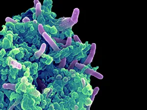
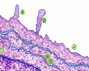
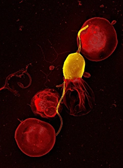
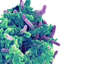
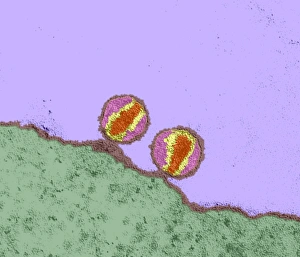
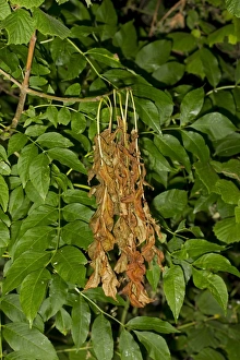
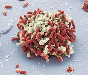
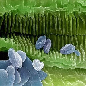
"Infecting the Microscopic World: Unveiling the Intricate Dance of Pathogens" Witness the captivating battle between bacteria and macrophages as they engage in a microscopic warfare. (Bacteria infecting a macrophage, SEM) Delve into the hidden realm of Hepatitis C viruses, observing their intricate structure under the powerful lens of TEM. Behold the cunning strategy employed by mouse malaria parasites as they invade their host's cells, captured through stunning SEM imagery. Explore the menacing Rift Valley fever virus, its sinister appearance revealed through TEM imaging techniques. Marvel at how bacteria infiltrate macrophages with astonishing precision, leaving us in awe of their adaptability and resilience. (Bacteria infecting a macrophage, SEM) Immerse yourself in an artistic representation showcasing bacteriophages - nature's very own viral assassins - engaged in their mission to eradicate harmful bacteria from our world. Reflect on humanity's crimes of ignorance as depicted in a poignant lithograph where people destroy disinfectants meant to protect them from harm. Transport yourself back to ancient times when Cerberus' foam infected Earth under Hercules' chains; an allegorical artwork that warns against unleashing chaos upon our planet. Observe Ash Dieback disease caused by Chalara fraxinea fungus as it ravages common ash trees' leaves, reminding us of nature's vulnerability to infections. Examine HIV-infected macrophages through SEM imagery – a visual reminder of the ongoing battle against this devastating virus that affects millions worldwide. Delve into the microscopic world once more and witness rust fungus infection up close.