
Immune Collection
"Unlocking the Power of Immunity: Exploring the Intricate World Within" Delving into the microscopic world


All Professionally Made to Order for Quick Shipping
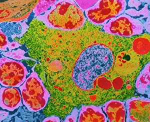
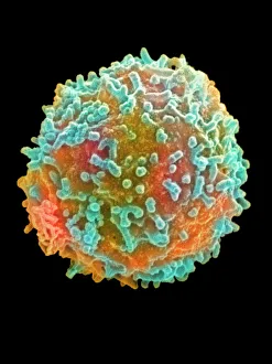
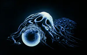
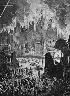
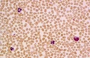
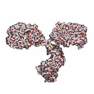
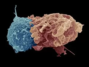
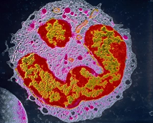
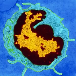
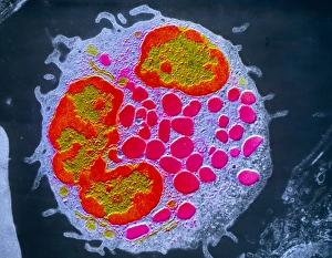
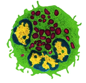
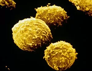
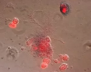
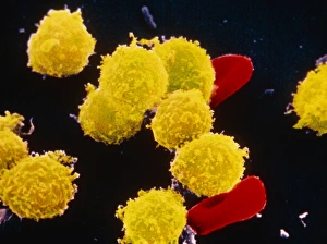
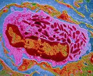
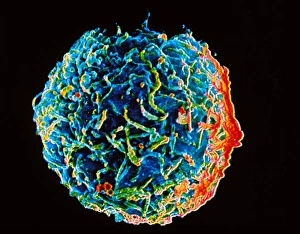
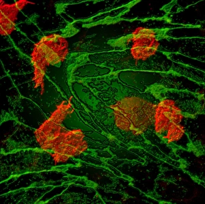
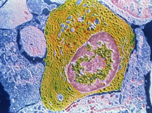
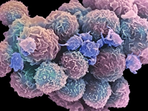
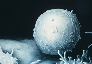
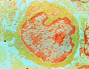
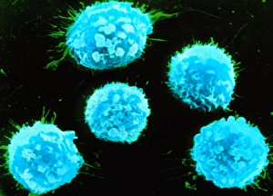
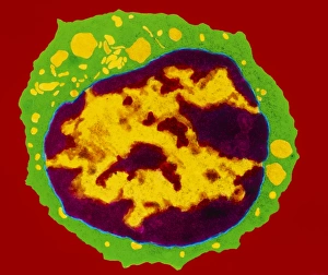
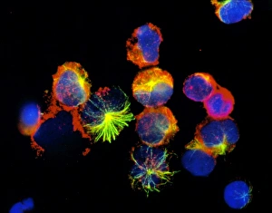
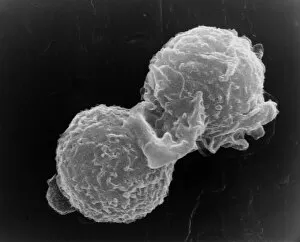
"Unlocking the Power of Immunity: Exploring the Intricate World Within" Delving into the microscopic world, a TEM image reveals the intricate structure of human macrophages, our immune system's frontline defenders. A mesmerizing Coloured SEM showcases a white blood cell, or lymphocyte, in action as it tirelessly patrols our body for invaders. In this modern-day fairy tale twist, "Sleeping Beauty - 6, " we witness how our immune system springs into action during slumber to protect us from harm. Witnessing an incredible feat of defense, a macrophage engulfs a bead with precision and efficiency, highlighting its role as an essential immune cell. The collaboration between white blood cells and platelets forms an unbreakable shield against pathogens that dare to threaten our well-being. Peering through the lens of a TEM microscope once again unveils the remarkable beauty and complexity of macrophage cells in their battle against infection. Reflecting on history's conquests and colonies in South America during the 16th century through an ancient lithographic map reminds us how immunity played a pivotal role in shaping civilizations. Breaking news alert. "New York Attacked" serves as a stark reminder that bolstering our immune defenses is crucial now more than ever before. Unexplained phenomena captured on film: The Hopkinsville UFO incident of 1955 raises questions about extraterrestrial encounters while reminding us to stay vigilant against unknown threats to our health. Zooming in on Immunoglobulin G antibody molecule C016 / 4462 reveals its vital role as one of nature's superheroes fighting off infections within our bodies' defense mechanisms. The smallpox scare lingers throughout history; here we see passengers being vaccinated before traveling to France—a testament to humanity's resilience when faced with deadly diseases.