
Iliac Artery Collection
The iliac artery is a vital component of the human circulatory system, playing a crucial role in maintaining proper blood flow throughout the body


All Professionally Made to Order for Quick Shipping
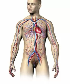
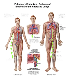
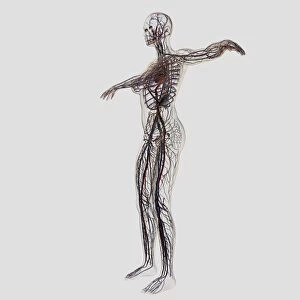
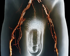
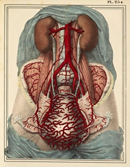
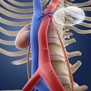
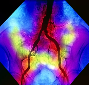
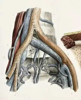
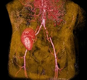
The iliac artery is a vital component of the human circulatory system, playing a crucial role in maintaining proper blood flow throughout the body. Located in the lower abdomen, this artery serves as a pathway for oxygenated blood to reach various organs and tissues. In cases of pulmonary embolism, where an embolus (a clot or foreign material) obstructs blood flow to the lungs, understanding the pathway through which it travels becomes essential. The iliac artery acts as one of these pathways, carrying deoxygenated blood from the lower limbs towards the heart and lungs. When examining the arteries and veins of the human body, it is fascinating to observe how they intricately intertwine with each other. A 3D CT scan provides us with a detailed view of these structures within a normal abdomen, allowing medical professionals to identify any abnormalities or blockages that may be present. For a more comprehensive understanding of our circulatory system's complexity, medical illustrations showcasing arteries, veins, and lymphatic systems in female midsections prove invaluable. These visual representations help us grasp how these networks function together seamlessly to ensure optimal health. Similarly enlightening are medical illustrations depicting male reproductive systems alongside their intricate network of veins and arteries. Such visuals shed light on how these components work harmoniously to support reproduction while also emphasizing their interconnectedness with other bodily systems. To truly appreciate just how remarkable our circulatory system is as a whole, full-length views displaying organs along with arteries and veins become necessary. These depictions provide an all-encompassing perspective on its vastness while highlighting its importance in sustaining life itself. Whether exploring male or female anatomy specifically or taking into account both genders collectively when studying arterial structures within our bodies remains crucial for comprehensive knowledge acquisition regarding this intricate web-like network that keeps us alive.