
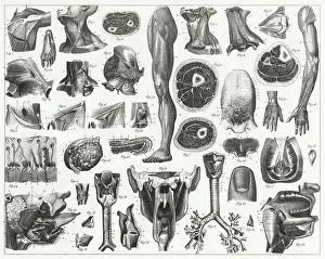
Human Cell Collection
The human cell, the building block of life, is a marvel of intricate design and functionality


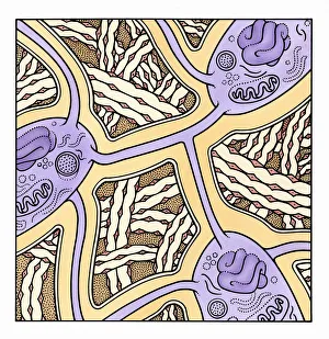
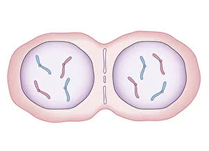
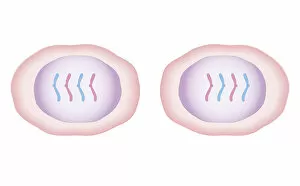
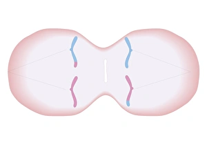
Digital illustration showing human cell division with chromosome making exact copy of itself and div
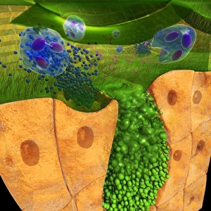
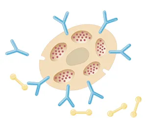
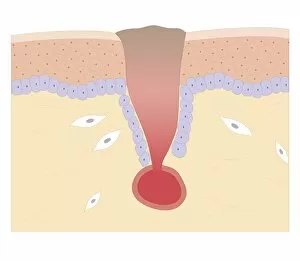
Digital cross section illustration of ciliate cell showing rhinovirus and antobodies in nasal cavity


All Professionally Made to Order for Quick Shipping
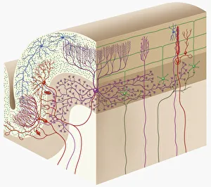
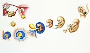
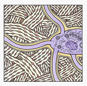
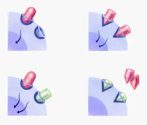
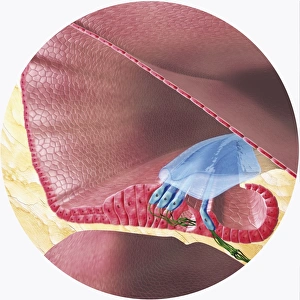
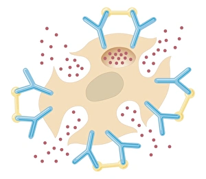
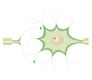
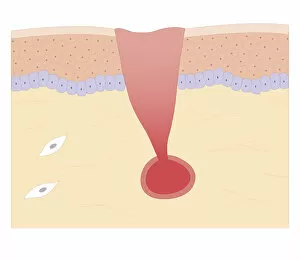
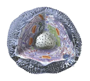
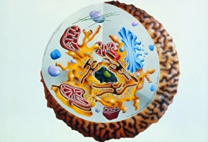
The human cell, the building block of life, is a marvel of intricate design and functionality. From the anatomy of organs engraving to the digital cross-section illustration of the human cerebellar cortex, we delve into the fascinating world within us. Witness the incredible journey from cells to a fully formed human being in a captivating drawing depicting the human life cycle. Explore further as an illustration showcases an astrocyte, one of many glial cells that support and nourish our neurons. Marvel at the formation of human bone through a mesmerizing digital illustration, highlighting its strength and resilience. Witness how chromosomes make exact copies during cell division, ensuring genetic continuity for future generations. Intriguingly, a digital cross-section illustration reveals ciliate cells with rhinovirus and antibodies in our nasal cavity – showcasing our body's defense mechanism against harmful invaders. Delve deeper into health matters as an illustration portrays osteoporosis affecting human bones, emphasizing the importance of maintaining bone density throughout life. Discover how different drugs affect cellular reactions through illustrations comparing agonist and antagonist drug responses. The complexity continues with a digital depiction of Corti organ found in our cochlea – responsible for hearing sounds that shape our perception of the world around us. Uncover penicillin's remarkable impact on bacteria through biomedical illustrations showcasing its entry into bacterium and subsequent destruction by interfering with essential chemicals needed for their survival. These captivating glimpses into various aspects biology remind us just how extraordinary each tiny unit is within ourselves - working tirelessly to sustain life and ensure our well-being.