Fraud Blocker Skip to main content

Histological Collection (page 5)

"Exploring the Intricacies of Histological Wonders: Unveiling the Hidden Beauty within Our Bodies" Step into the fascinating world of histology

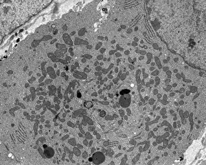
Background imageHistological Collection: Pancreatic acinar cell, TEM

Pancreatic acinar cell, TEM
Pancreatic acinar cell. Transmission electron micrograph (TEM) of a section through an enzyme-secreting acinar cell in the human pancreas, showing the nucleus (dark purple, centre)

Background imageHistological Collection: Activated plasma cell, TEM

Activated plasma cell, TEM
Activated plasma cell, coloured transmission electron micrograph (TEM). Plasma cells are derived from stimulated B lymphocytes (white blood cells)

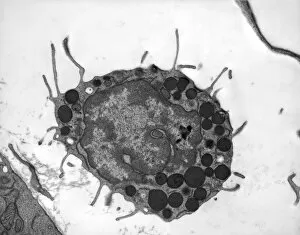
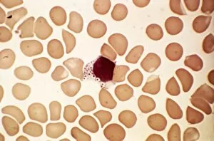
Background imageHistological Collection: Basophil white blood cell, TEM

Basophil white blood cell, TEM
Basophil white blood cell. Transmission electron micrograph (TEM) of section through a circulating basophil cell. Basophils are the smallest and least common of the white blood cells

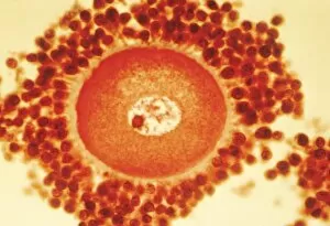
Background imageHistological Collection: Oocyte before ovulation, light micrograph

Oocyte before ovulation, light micrograph
Oocyte before ovulation. Light micrograph of a section through tissue from an ovary, showing an oocyte two days before ovulation

Background imageHistological Collection: Lymphoblastic sarcoma, light micrograph

Lymphoblastic sarcoma, light micrograph
Lymphoblastic sarcoma. Light micrograph of blood cells from bone marrow in a case of lymphoblastic sarcoma. These small lymphoblasts (dark red) characteristically have conspicuous nucleoli

Background imageHistological Collection: Plasmocyte blood cell, light micrograph

Plasmocyte blood cell, light micrograph. Plasmocytes, also called plasma cells, are derived from stimulated B lymphocytes (white blood cells)

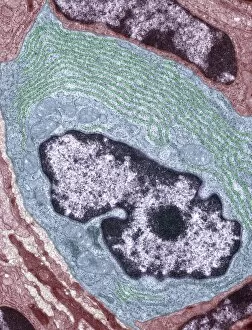
Background imageHistological Collection: Myenteric nerve plexus, TEM

Myenteric nerve plexus, TEM
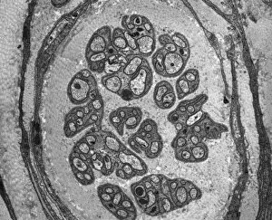
Myenteric nerve plexus. Transmission electron micrograph (TEM) of a section through a bundle of unmyelinated nerve fibres in the myenteric nerve plexus of the intestinal tract

Background imageHistological Collection: Multiple myeloma, light micrograph

Multiple myeloma, light micrograph
Multiple myeloma. Light micrograph of dystrophy (abnormality) of plasmocyte blood cells from bone marrow in a case of multiple myeloma, also known as Kahlers disease or plasma cell myeloma

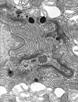
Background imageHistological Collection: Golgi membranes, TEM

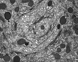
Golgi membranes, TEM
Golgi membranes. Transmission electron micrograph (TEM) of a section through a cell, showing the membranes (dark lines) of the Golgi apparatus

Background imageHistological Collection: Lymphoplasmacytic lymphoma, micrograph

Lymphoplasmacytic lymphoma, micrograph
Lymphoplasmacytic lymphoma. Light micrograph of a mixture of small lymphocytes and plasma cells from bone marrow in a case of lymphoplasmacytic lymphoma, also known as Waldenstroms macroglobulinemia

Background imageHistological Collection: Ovarian follicle tissue, light micrograph

Ovarian follicle tissue, light micrograph
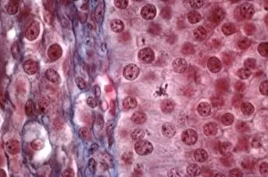
Ovarian follicle tissue. Light micrograph of a section through tissue from an ovarian follicle, showing a boundary between two layers

Background imageHistological Collection: Goblet cell, TEM

Goblet cell, TEM
Goblet cell. Transmission electron micrograph (TEM) of a section through a goblet cell in the lining (epithelium, top) of the small intestine

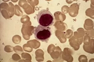
Background imageHistological Collection: Hairy cell leukaemia, light micrograph

Hairy cell leukaemia, light micrograph
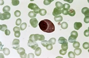
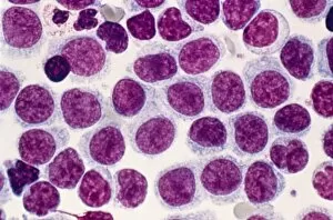
Hairy cell leukaemia. Light micrograph of blood cells in a case of hairy cell leukaemia. This sample, treated with May-Grunwald-Giemsa (MGG) stain

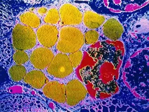
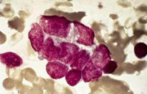
Background imageHistological Collection: Mast cells, light micrograph

Mast cells, light micrograph
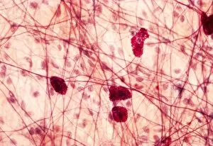
Mast cells. Light micrograph of several mast cells (pink rounded objects). These leucocytes (white blood cells) contain the chemical mediators histamine, serotonin and heparin

Background imageHistological Collection: Smooth muscle of gut wall, TEM

Smooth muscle of gut wall, TEM
Smooth muscle of gut wall. Transmission electron micrograph (TEM) of a section through the outermost wall of the intestine

Background imageHistological Collection: Lymphocyte white blood cell, TEM

Lymphocyte white blood cell, TEM
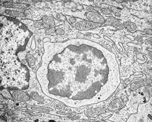
Lymphocyte white blood cell. Transmission electron micrograph (TEM) of a section through lymphocyte, showing the nucleus (large, round, centre), endoplasmic reticulum (ER)

Background imageHistological Collection: Cervix, light micrograph

Cervix, light micrograph
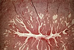
Cervix. Light micrograph of a section through the mucosa of the cervix, the neck of the uterus. This is the endocervix, which borders the uterus. It is lined with columnar epithelial cells

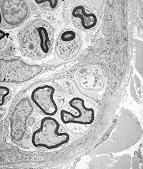
Background imageHistological Collection: Unmyelinated nerve, TEM

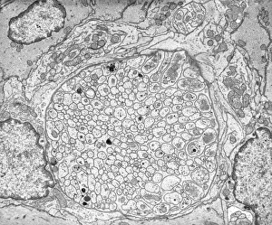
Unmyelinated nerve, TEM
Unmyelinated nerve. Transmission electron micrograph (TEM) of a section through axon (nerve fibre) bundles of unmyelinated nerves

Background imageHistological Collection: Steroid secreting cell, TEM

Steroid secreting cell, TEM
Steroid secreting cell. Transmission electron micrograph (TEM) of a section through a cell involved with steroid synthesis and secretion (steroidogenesis)

Background imageHistological Collection: Acute erythroid leukaemia, micrograph

Acute erythroid leukaemia, micrograph
Acute erythroid leukaemia. Light micrograph of blood cells from bone marrow in a case of acute erythroid leukaemia. The cells include dystrophic (degenerated) erythroblasts (nuclei stained dark red)

Background imageHistological Collection: Enterocyte, TEM

Enterocyte, TEM
Enterocyte. Transmission electron micrograph (TEM) of a section through the cytoplasm and part of the nucleus of an enterocyte cell located in a crypt of Lieberkuhn of the small intestine

Background imageHistological Collection: Acute lymphoblastic leukaemia, micrograph

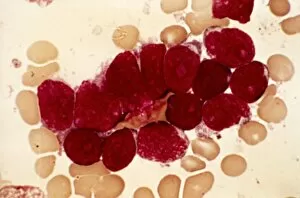
Acute lymphoblastic leukaemia, micrograph
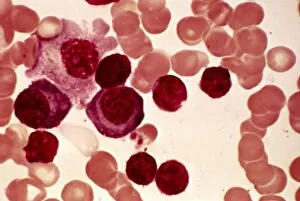
Acute lymphoblastic leukaemia. Light micrograph of blood cells from bone marrow in a case of acute lymphoblastic leukaemia. Leukaemia is a cancer where certain blood cells form in excess

Background imageHistological Collection: Malignant histiocytosis, light micrograph

Malignant histiocytosis, light micrograph
Malignant histiocytosis. Light micrograph of blood cells in a case of malignant histiocytosis. Hystiocytosis is an excess number of histiocytes

Background imageHistological Collection: Islet of Langerhans, TEM

Islet of Langerhans, TEM
Islet of Langerhans. Transmission electron micrograph (TEM) of a section through cells clustered in an islet of Langerhans in the pancreas

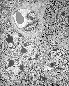
Background imageHistological Collection: Capillary, TEM

Capillary, TEM
Capillary. Transmission electron micrograph (TEM) of a section through a capillary, showing two red blood cells (erythrocytes, black) in its interior

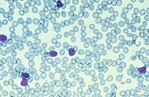
Background imageHistological Collection: Blood cells, light micrograph

Blood cells, light micrograph
Blood cells. Light micrograph of red blood cells (erythrocytes, light blue) and white blood cells (leucocytes, nuclei stained purple)

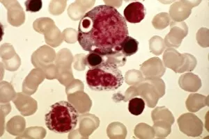
Background imageHistological Collection: Chediak-Higashi syndrome, micrograph

Chediak-Higashi syndrome, micrograph
Chediak-Higashi syndrome. Light micrograph of blood cells in a case of Chediak-Higashi syndrome. The cells include neutrophil white blood cells with abnormal granules

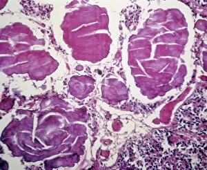
Background imageHistological Collection: Metastatic breast cancer, micrograph

Metastatic breast cancer, micrograph
Metastatic breast cancer. Light micrograph of blood cells in a case of metastatic (secondary) medullary breast cancer. Medullary breast cancer is a rare form of breast cancer that is diagnosed by

Background imageHistological Collection: Dividing cell, TEM

Dividing cell, TEM
Dividing cell. Transmission electron micrograph (TEM) of a section through a cell dividing through the process of mitosis

Background imageHistological Collection: Metastatic lung cancer, micrograph

Metastatic lung cancer, micrograph
Metastatic lung cancer. Light micrograph of blood cells in a case of metastatic (secondary) medullary lung cancer. Medullary cancer (carcinoma)

Background imageHistological Collection: Pineal gland, light micrograph

Pineal gland, light micrograph. The pineal gland is a small endocrine gland located near to the centre of the brain, between the two hemispheres

Background imageHistological Collection: Myelinated nerve tissue, TEM

Myelinated nerve tissue, TEM
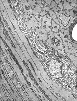
Myelinated nerve tissue. Transmission electron micrograph (TEM) of a section through myelinated nerve fibres (axons). Each axon is coated with many layers of myelin

Background imageHistological Collection: Alders anomaly, light micrograph

Alders anomaly, light micrograph
Alders anomaly. Light micrograph of an abnormal lymphocyte white blood cell (centre) from a blood sample in a case of Alders anomaly

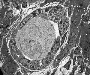
Background imageHistological Collection: Ovarian follicle, TEM

Ovarian follicle, TEM
Ovarian follicle. Transmission electron micrograph (TEM) of a section through an ovary, showing a primary follicle. Primary follicles contain a central oocyte (female germ cell)


All Professionally Made to Order for Quick Shipping

"Exploring the Intricacies of Histological Wonders: Unveiling the Hidden Beauty within Our Bodies" Step into the fascinating world of histology, where microscopic wonders come to life. Delve into Santiago Ramon Y Cajal's masterpiece, as his cortical grey matter schema unveils the intricate network that forms our brain's command center. Witness the mesmerizing complexity of a mammalian retina through a histological diagram, revealing how light is transformed into vision. Journey deeper into our cerebellum tissue, as a captivating light micrograph showcases its unique structure and function. Marvel at Ramon Y Cajal's genius once again as he unravels the secrets of synapse nerve junctions through an astonishing TEM image. Venture further into uncharted territory with a glimpse of hippocampus brain tissue, where memories are formed and stored. Observe Purkinje nerve cells in the cerebellum, their elegant arrangement hinting at their crucial role in coordinating movement. Witness nature's precision with kidney tubules in section; these delicate structures filter waste from our bloodstreams tirelessly. Explore human brain microscope slides and be awestruck by its intricate architecture - billions of neurons working harmoniously to shape who we are. Peer closely at rough endoplasmic reticulum through TEM imagery; this cellular powerhouse orchestrates protein synthesis within each living cell. Immerse yourself in glial stem cell culture under a light microscope - witness new life being nurtured for potential regeneration. Finally, marvel at the intricacy of brain tissue blood supply - countless vessels ensuring oxygen and nutrients reach every corner of this remarkable organ. Histological wonders unlock hidden beauty within us all – reminding us that even on a microscopic level, there is awe-inspiring complexity waiting to be discovered.