
Hemagglutinin Collection
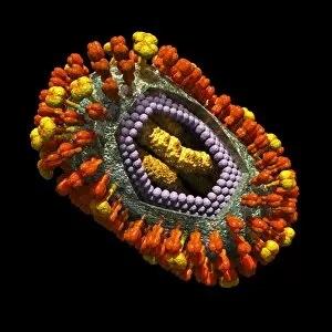
Hemagglutinin, a key protein found on the surface of influenza viruses, plays a crucial role in their ability to infect and spread


All Professionally Made to Order for Quick Shipping
Hemagglutinin, a key protein found on the surface of influenza viruses, plays a crucial role in their ability to infect and spread. This microscopic view showcases the H5N1 virus alongside red and white blood cells, highlighting its potential impact on human health. In another image, a black swarm of H5N1 avian flu viruses is under attack by antibodies, illustrating our immune system's defense mechanism against these harmful pathogens. The TEM images of avian influenza virus H7N9 further emphasize the intricate structure of this viral strain. With each magnified detail captured in C016 / 6294, C016 / 6293, and C016 / 6292, we gain insight into its morphology and composition. Similarly, TEM images labeled as C015 / 8800, C015 / 8799, and C015 / 8797 depict different strains of avian influenza virus with distinct characteristics worth exploring. Influenza viruses are notorious for causing seasonal outbreaks worldwide; hence understanding their structure becomes imperative in combating them effectively. Illustrations like artwork C018/0735 showcase an artistic representation of an influenza virus while providing insights into its inner workings that contribute to infection. Artworks labeled as C016/8349, C016/8348, andC016/8347 further highlight various aspects such as viral replication or host cell interaction. Through these captivating visuals capturing hemagglutinin's presence within diverse strains of influenza viruses - from H5N1 to H7N9 - scientists can delve deeper into studying their behavior and developing effective preventive measures such as vaccines or antiviral drugs that target this critical protein component.