
Hela Cell Collection
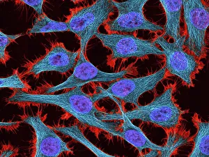
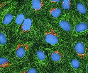
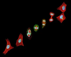
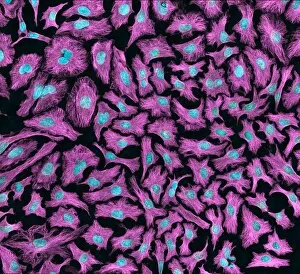
"HeLa Cells: A Window into Cellular Life and Death" The captivating world of HeLa cells, as revealed through light and scanning electron micrographs


All Professionally Made to Order for Quick Shipping
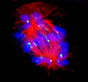
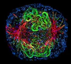
"HeLa Cells: A Window into Cellular Life and Death" The captivating world of HeLa cells, as revealed through light and scanning electron micrographs, offers a glimpse into the intricate processes of mitosis and cell division. In the light micrograph C017 / 8299, we witness the vibrant beauty of HeLa cells in their natural state. The adjacent image, C017 / 8298, showcases these dividing cells in action during mitosis. Moving on to scanning electron microscopy (SEM), the SEM images C014 / 0371, C014 / 0366, C014 / 0369, and C017 / 8305 provide an astonishingly detailed view of HeLa cells' structural complexity. These images highlight the delicate intricacies that make up these remarkable cellular entities. However, not all is eternal within this microscopic realm. As depicted in SEM image C017 / 8304, we observe a poignant moment where a HeLa cell reaches its demise—a reminder that life's cycle encompasses both creation and decay. Returning to light microscopy with images C013 / 4774 and C013/4773 showcasing more instances of mitosis among HeLa cells—these snapshots serve as reminders that life perpetually moves forward. Mitotic events are crucial for growth and regeneration within our bodies. HeLa cells have revolutionized medical research since their discovery in Henrietta Lacks' cervical tumor back in the early '50s. Their unique ability to replicate indefinitely has paved new avenues for scientific breakthroughs across various disciplines. Exploring these mesmerizing glimpses into the world of HeLa cells reveals not only their stunning visual appeal but also sheds light on fundamental biological processes such as mitosis and cellular death. These tiny powerhouses continue to shape our understanding of life at its most basic level while leaving us awestruck by their resilience and adaptability.