
Granule Collection (page 2)
Granules are tiny particles that can be found in various contexts, from the microscopic world of cells to the vast landscapes of nature


All Professionally Made to Order for Quick Shipping
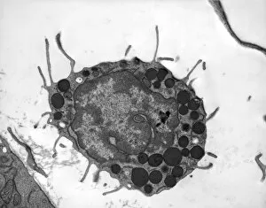
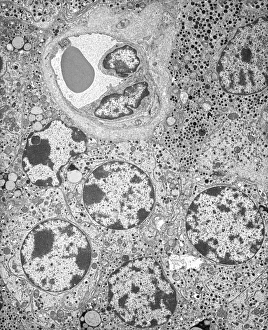
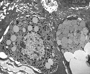
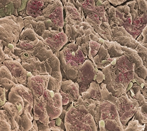
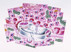
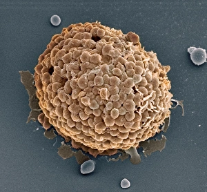
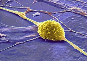
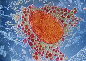
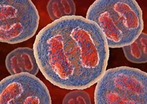
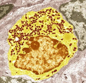
Granules are tiny particles that can be found in various contexts, from the microscopic world of cells to the vast landscapes of nature. In the realm of biology, granules play a crucial role in the functioning of our immune system. Basophil white blood cells, for instance, contain granules that release histamine and other chemicals involved in allergic reactions. Examining a blood cell under a microscope reveals intriguing details like Dohle bodies - small blue-stained granular structures within these vital carriers of life. Such micrographs offer glimpses into the intricate workings of our bodies on a cellular level. Shifting gears to industry, we find an employee diligently working at Krastsvetmet non-ferrous metals plant in Krasnoyarsk. Amidst this bustling environment where metals are transformed and shaped, one can imagine countless granules being manipulated and processed with precision. On another note entirely, let's zoom into everyday objects that often go unnoticed. A close-up detail captures an espresso maker and coffee beans - each bean representing a tiny granule filled with flavor potential waiting to be released by hot water coursing through it. Nature too has its own way of showcasing the beauty of granules. A scenic view unfolds before us as we gaze upon Northumberland's lighthouse standing tall against crashing waves. The surrounding landscape is adorned with countless grains of sand - each grain formed over time by erosion and weathering processes. As we explore further through pictures numbered 11014611 to 11014602, we encounter diverse scenes: from serene seascapes to vibrant cityscapes; from breathtaking sunsets to architectural marvels; all reminding us that even on larger scales, granular elements contribute significantly to our visual experiences. Whether they reside within our bodies or shape our surroundings both industrially and naturally, granules hold immense significance.