
Granular Layer Collection
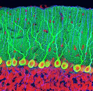
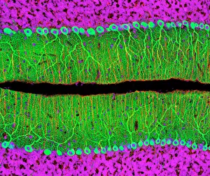
The granular layer, found in the cerebellum, is a fascinating and intricate structure that plays a crucial role in our motor coordination


All Professionally Made to Order for Quick Shipping
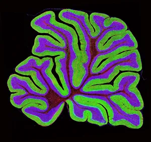
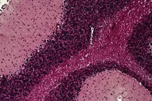
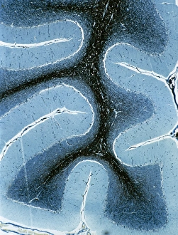
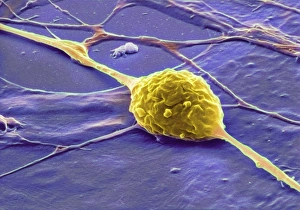
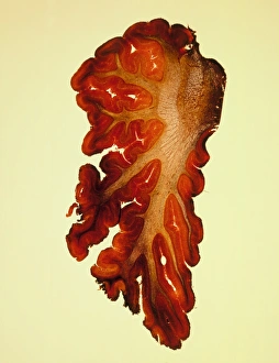
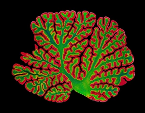
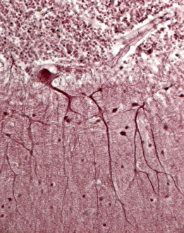
The granular layer, found in the cerebellum, is a fascinating and intricate structure that plays a crucial role in our motor coordination. Purkinje nerve cells, located within this layer, are key players in transmitting information from the cerebellum to other parts of the brain. These specialized cells have an elaborate branching pattern with numerous dendrites that receive signals from various sources. When observed under a light microscope, the cerebellum's granular layer reveals its complex organization. The tissue appears densely packed with small granule cells, giving it its distinctive appearance. This microscopic view showcases the intricacy and interconnectedness of these vital neural components. In contrast to the neighboring cerebrum's structure seen through another light micrograph, where different regions are responsible for higher cognitive functions such as memory and language processing, the cerebellum focuses on fine-tuning movements and maintaining balance. Within this granular layer lies both cortex and medulla regions of the cerebellum. This layered composition further emphasizes its complexity and highlights how each component contributes to overall motor control. As we zoom closer into this tissue using high-resolution microscopy techniques, Purkinje nerve cells become more apparent. Their distinct morphology stands out against the surrounding granule cells - large cell bodies with extensive dendritic trees reaching towards other layers of the cerebellar cortex. Through multiple light micrographs capturing different sections of this remarkable tissue sample, we gain insight into how these Purkinje nerve cells populate throughout the granular layer. Their abundance signifies their importance in relaying information effectively within this region.