
Gram Positive Collection
Gram-positive bacteria are a diverse group of microorganisms that can be found in various environments


All Professionally Made to Order for Quick Shipping
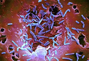
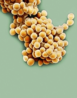
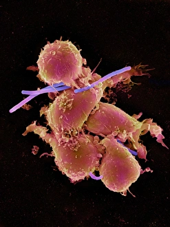
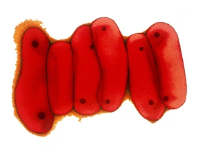
Gram-positive bacteria are a diverse group of microorganisms that can be found in various environments. These fascinating organisms come in different shapes and sizes, as depicted in stunning artwork and scanning electron microscope (SEM) images. One captivating image shows a neutrophil engulfing MRSA, a type of gram-positive bacterium known for its resistance to antibiotics. This SEM image, labeled C018 / 8596, highlights the battle between the immune system and harmful bacteria. Another SEM image showcases tuberculosis bacteria, which belong to the Mycobacterium genus. These rod-shaped bacilli are responsible for causing one of humanity's oldest infectious diseases. Mycobacterium chelonae is another intriguing member of the gram-positive family. An SEM image reveals its unique structure and features (C014 / 0631), providing valuable insights into this bacterial species. In contrast to pathogenic bacteria, some gram-positive organisms have beneficial roles. Lactobacillus bacteria play an essential role in maintaining our gut health. A mesmerizing SEM image captures their intricate details up close. The Bacillus genus encompasses several gram-positive bacteria with distinct characteristics. Their varied shapes and arrangements make them visually striking under microscopic examination. Staphylococcus aureus is a well-known gram-positive bacterium notorious for causing infections in humans. Its spherical shape is beautifully captured in an SEM image labeled C017 / 7137, showcasing its surface features. When MRSA encounters neutrophils – our body's first line of defense against infection – intense battles occur within our bodies' microscopic realm. Another compelling SEM image (C018 / 8601) portrays this interaction between MRSA and a dead neutrophil. But not all interactions end tragically; some neutrophils successfully engulf MRSA to neutralize the threat they pose. The captivating SEM image C018 / 8597 illustrates this victorious moment when our immune system overpowers harmful pathogens like MRSA.