
Glial Collection
"Unveiling the Intricacies of Glial Cells in the Hippocampus and Retina" Delving into the depths of brain tissue, we encounter a fascinating world known as glial cells


All Professionally Made to Order for Quick Shipping
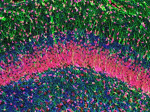
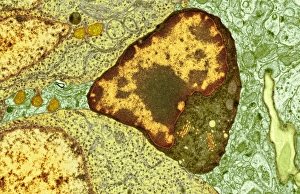
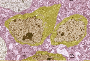
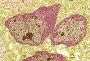
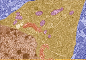
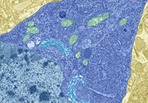
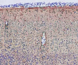
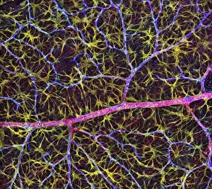
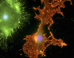
"Unveiling the Intricacies of Glial Cells in the Hippocampus and Retina" Delving into the depths of brain tissue, we encounter a fascinating world known as glial cells. These unsung heroes play a crucial role in supporting and nourishing our neurons, ensuring optimal brain function. In an intricate illustration, we witness the presence of astrocyte human glial cells. With their star-like shape and numerous branches, they form an extensive network throughout the hippocampus region. Their vital task involves regulating neurotransmitter levels, maintaining chemical balance, and providing structural support to neurons. Through electron microscopy images labeled TEM C014 / 0358 and TEM C014 / 0359, we gain a closer look at these remarkable brain cells. The high-resolution snapshots reveal their complex structure with precision – each cell meticulously intertwined with others like puzzle pieces forming an elaborate tapestry within our minds. Further exploration through TEM C013 / 4801 and TEM C013 / 4800 showcases additional brain cells working harmoniously together. These microscopic wonders collaborate tirelessly to ensure smooth communication between neurons while also protecting them from harm. As we continue our journey through this captivating realm, another image labeled TEM C013 / 4799 captures yet another glimpse into the mesmerizing intricacy of these glial cells. Their interconnectedness becomes even more apparent as they weave around one another like dancers performing an elegant ballet on nature's grandest stage – our brains. Within this vast landscape lies not only the hippocampus but also retina blood vessels intertwined with nerve cells. This unique combination is beautifully depicted in multiple illustrations showcasing both structures coexisting harmoniously within delicate tissues. Returning once again to explore hippocampus brain tissue reveals its significance as a hub for memory formation and spatial navigation. Herein lies evidence of how glial cells contribute to these cognitive processes by nurturing neuronal connections essential for learning and memory retention.