Fraud Blocker Skip to main content

Germ Cell Collection

"Germ Cell: Unveiling the Intricacies of Life's Building Blocks" In the realm of cellular biology

Background imageGerm Cell Collection: Secondary heart cancer, light micrograph F005 / 6069

Secondary heart cancer, light micrograph F005 / 6069
Secondary heart cancer. Light micrograph of a section through cardiac muscle showing a metastatic seminoma. This is a germ cell tumour that originated in the testis

Background imageGerm Cell Collection: Oocyte, light micrograph

Oocyte, light micrograph
Oocyte. Light micrograph of a section through an oocyte within a early antral follicle in an ovary. Oocytes are immature ova, or egg cells

Background imageGerm Cell Collection: Roundworm germ cells, light micrograph C016 / 9538

Roundworm germ cells, light micrograph C016 / 9538
Roundworm germ cells. Light micrograph of germ cells from a roundworm (Ascaris sp.), undergoing mitosis (nuclear division)

Background imageGerm Cell Collection: Ovarian follicle, TEM

Ovarian follicle, TEM
Ovarian follicle. Transmission electron micrograph (TEM) of a section through an ovary, showing a primary follicle. Primary follicles contain a central oocyte (female germ cell)

Background imageGerm Cell Collection: Ovarian primordial follicles, micrograph C016 / 0518

Ovarian primordial follicles, micrograph C016 / 0518
Ovarian primordial follicles. Light micrograph of a section through the cortex of a neonatal ovary, showing clusters of primordial follicles each containing a single oocyte (egg cell)

Background imageGerm Cell Collection: Oocytes, illustration C018 / 0808

Oocytes, illustration C018 / 0808
Oocytes, illustration. Oocytes are immature egg cells, the female gametes (sex cells). Oocytes are encased in a glycoprotein shell called the zona pellucida that prevents more than one sperm

Background imageGerm Cell Collection: Dermoid ovarian cyst C015 / 6910

Dermoid ovarian cyst C015 / 6910
Dermoid ovarian cyst. Light micrograph of a section through an ovary with a dermoid, or teratoma, ovarian cyst. This is a benign (non-cancerous) cyst that arises from germ (sex) cells

Background imageGerm Cell Collection: Dermoid ovarian cyst C015 / 6056

Dermoid ovarian cyst C015 / 6056
Dermoid ovarian cyst. Light micrograph of a section through an ovary with a dermoid, or teratoma, ovarian cyst. This is a benign (non-cancerous) cyst that arises from germ (sex) cells

Background imageGerm Cell Collection: Dermoid ovarian cyst C015 / 6402

Dermoid ovarian cyst C015 / 6402
Dermoid ovarian cyst. Light micrograph of a section through an ovary with a dermoid, or teratoma, ovarian cyst. This is a benign (non-cancerous) cyst that arises from germ (sex) cells

Background imageGerm Cell Collection: Fertilisation

Fertilisation, computer artwork. Sperm fertilising an egg

Background imageGerm Cell Collection: ZP3 glycoprotein molecule

ZP3 glycoprotein molecule
ZP3 glycoprotein. Computer artwork of a molecule of ZP3 (zona pellucida glycoprotein 3), a sperm receptor in the external membrane (zona pellucida) of a female germ cell (egg)


All Professionally Made to Order for Quick Shipping

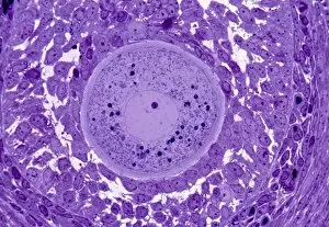
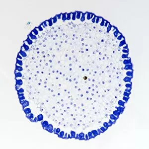
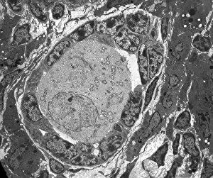
"Germ Cell: Unveiling the Intricacies of Life's Building Blocks" In the realm of cellular biology, germ cells hold a profound significance as they play a pivotal role in the perpetuation and continuation of life. These enigmatic entities have captivated scientists with their unique characteristics and diverse functions. From battling secondary heart cancer to nurturing oocytes, from testicular cancer to roundworm germ cells, each microscopic glimpse offers a window into this fascinating world. Under the watchful eye of light micrographs F005/6069, we witness the intricate dance between germ cells and secondary heart cancer. This visual revelation serves as a reminder of both resilience and vulnerability within our bodies. Delving deeper into this captivating subject matter, light micrographs unveil another facet - oocytes. These precious gems hold immense potential for future generations, representing hope and continuity amidst the complex tapestry of life. Testicular cancer takes center stage under yet another light micrograph's lens. The delicate balance between health and disease is laid bare before us, urging researchers to unravel its mysteries in pursuit of effective treatments that can save lives. Roundworms may seem insignificant at first glance; however, their germ cells offer invaluable insights into fundamental biological processes. Light micrographs C016/9541 & C016/9538 reveal these tiny organisms' hidden secrets – an intricate web connecting reproduction with survival. Apoptosis cell death emerges as an essential component in understanding germ cell dynamics through TEM imagery. This phenomenon showcases nature's way of maintaining equilibrium by eliminating damaged or unnecessary cells while paving the path for new beginnings. TEM images provide an even closer look at human spermatids - remarkable structures on their journey towards maturity. Within these minute forms lies tremendous potential for creating life itself; it is awe-inspiring how such minuscule entities possess such extraordinary power. The battle against testicular cancer continues under TEM scrutiny as researchers strive to comprehend its intricacies at a cellular level.