Fraud Blocker Skip to main content

Genetic Collection (page 9)

"Unlocking the Secrets: Exploring the Fascinating World of Genetics" In this captivating journey, we delve into the intricate realm of genetics

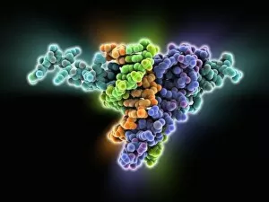
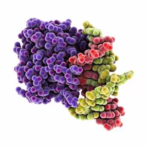
Background imageGenetic Collection: Metal-binding protein bound to DNA

Metal-binding protein bound to DNA. Molecular model of the bacterial metal-binding protein NikR (bottom) bound to a strand of DNA (top, helical, deoxyribonucleic acid)

Background imageGenetic Collection: Haematopoietic stem cells, artwork

Haematopoietic stem cells, artwork
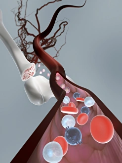
Haematopoietic stem cells. Cutaway computer artwork showing white blood cells (leucocytes, white, round), red blood cells (erythrocytes, red) and haematopoietic stem cells (HSCs)

Background imageGenetic Collection: Fertilisation, artwork C016 / 7516

Fertilisation, artwork C016 / 7516
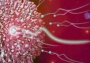
Fertilisation. Cutaway computer artwork of a sperm cell (spermatozoa) penetrating an egg (large). The nucleus of the sperm cell will fuse with the egg and its DNA (deoxyribonucleic acid)

Background imageGenetic Collection: Spider silk research C016 / 9552

Spider silk research C016 / 9552
Spider silk research. Golden silk orb-weaver spider in a laboratory being used for research on spider silk proteins. A thin strand of silk is visible emerging from the spiders spinnerets (centre)

Background imageGenetic Collection: DNA packaging, artwork C016 / 7517

DNA packaging, artwork C016 / 7517
DNA packaging. Computer artwork showing how DNA (deoxyribonucleic acid) is packaged within cells. Two DNA strands, consisting of a sugar-phosphate backbone attached to nucleotide bases

Background imageGenetic Collection: Roundworm germ cells, light micrograph C016 / 9538

Roundworm germ cells, light micrograph C016 / 9538
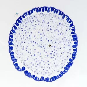
Roundworm germ cells. Light micrograph of germ cells from a roundworm (Ascaris sp.), undergoing mitosis (nuclear division)

Background imageGenetic Collection: Plant genetics research C016 / 9539

Plant genetics research C016 / 9539
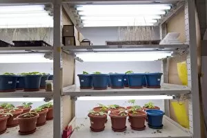
Plant genetics research. Morning glory (Ipomoea cairica) plant seedlings in an incubator at a genetics research laboratory

Background imageGenetic Collection: Genetic fingerprints, conceptual artwork C016 / 7521

Genetic fingerprints, conceptual artwork C016 / 7521
Genetic fingerprints, conceptual computer artwork

Background imageGenetic Collection: Fertilisation, artwork C016 / 7515

Fertilisation, artwork C016 / 7515
Fertilisation. Computer artwork of a sperm cell (spermatozoa) penetrating an egg (large)

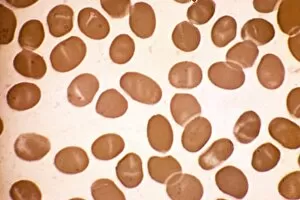
Background imageGenetic Collection: Elliptocytosis, light micrograph

Elliptocytosis, light micrograph
Elliptocytosis. Light micrograph of red blood cells in a case of elliptocytosis. Red blood cells (erythrocytes) carry oxygen and carbon dioxide to and from body tissues

Background imageGenetic Collection: Genomic HIV-RNA duplex

Genomic HIV-RNA duplex, molecular model. This structure shows the dimerization initiation site of genomic HIV-1 with RNA (ribonucleic acid)

Background imageGenetic Collection: Genetically modified tomato, artwork

Genetically modified tomato, artwork
Genetically modified tomato. Conceptual artwork representing genetic modification, showing a lemon (yellow) partially depicted on a tomato

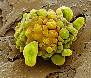
Background imageGenetic Collection: Stem cell dying, SEM

Stem cell dying, SEM
Stem cell dying. Coloured scanning electron micrograph (SEM) of a stem cell undergoing apoptosis, or programmed cell death. Apoptosis occurs when a cell becomes old or damaged

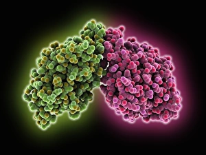
Background imageGenetic Collection: Chromosome segregation protein

Chromosome segregation protein, molecular model. This proteins function is to aid the process of chromosome segregation during cell division and replication

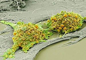
Background imageGenetic Collection: Mesenchymal stem cells, SEM

Mesenchymal stem cells, SEM
Mesenchymal stem cells. Coloured scanning electron micrograph (SEM) of two human mesenchymal stem cells (MSCs). MSCs are multipotent stromal (connective tissue)

Background imageGenetic Collection: Svalbard Global Seed Vault, artwork

Svalbard Global Seed Vault, artwork
Svalbard Global Seed Vault, conceptual artwork. Polar bear walking on snow and ice above a vault containing fruits. This represents the Svalbard Global Seed Vault, constructed between 2006 and 2008

Background imageGenetic Collection: Genetically modified grapes, artwork

Genetically modified grapes, artwork
Genetically modified grapes. Conceptual artwork of grapes in the double helix shape of DNA (deoxyribonucleic acid) on a vine. This represents concepts such as the genetic modification of food crops

Background imageGenetic Collection: Malignant histiocytosis, light micrograph

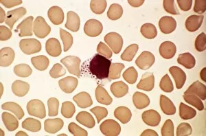
Malignant histiocytosis, light micrograph
Malignant histiocytosis. Light micrograph of blood cells in a case of malignant histiocytosis. Hystiocytosis is an excess number of histiocytes

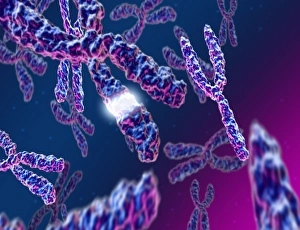
Background imageGenetic Collection: Male XY chromosomes, artwork

Male XY chromosomes, artwork. The Y chromosome (right) is found in males and is shorter than the X chromosome (upper left)

Background imageGenetic Collection: Chediak-Higashi syndrome, micrograph

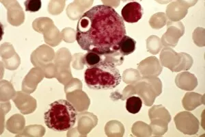
Chediak-Higashi syndrome, micrograph
Chediak-Higashi syndrome. Light micrograph of blood cells in a case of Chediak-Higashi syndrome. The cells include neutrophil white blood cells with abnormal granules

Background imageGenetic Collection: Thymidylic acid-ribonuclease A complex

Thymidylic acid-ribonuclease A complex. Molecular model of a thymidylic acid tetramer (blue) in complex with ribonuclease A (red)

Background imageGenetic Collection: Alders anomaly, light micrograph

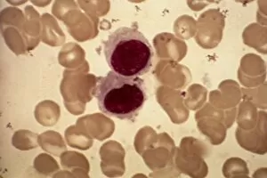
Alders anomaly, light micrograph
Alders anomaly. Light micrograph of an abnormal lymphocyte white blood cell (centre) from a blood sample in a case of Alders anomaly

Background imageGenetic Collection: Polycystic kidney

Polycystic kidney. Gross specimen of a polycystic kidney, where most of the tissue of the kidneys has been replaced by cysts

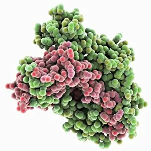
Background imageGenetic Collection: Exosome complex, molecular model

Exosome complex, molecular model. This multi-protein complex functions to break up strands of RNA (ribonucleic acid, pink) during biochemical processes

Background imageGenetic Collection: MyoD muscle protein-DNA complex

MyoD muscle protein-DNA complex. Molecular model of the MyoD muscle protein (green and blue) bound to a strand of DNA (deoxyribonucleic acid, orange and green)

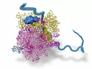
Background imageGenetic Collection: Bacterial ribosome and protein synthesis

Bacterial ribosome and protein synthesis. Molecular model showing a bacterial ribosome reading an mRNA (messenger ribonucleic acid) strand (blue) and synthesising a protein

Background imageGenetic Collection: Tumour suppressor protein and DNA C016 / 6264

Tumour suppressor protein and DNA C016 / 6264
Tumour suppressor protein and DNA. Computer artwork showing a molecule of the tumour suppressor protein p53 (blue and green) bound to a strand of DNA (deoxyribonucleic acid, grey)

Background imageGenetic Collection: Oncogenes, artwork C016 / 6262

Oncogenes, artwork C016 / 6262
Oncogenes. Computer artwork comparing the DNA (deoxyribonucleic acid, coiled strand) of a normal cell (top) with that of a cancer cell (bottom)

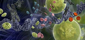
Background imageGenetic Collection: Drug effect on viruses, conceptual image C016 / 6253

Drug effect on viruses, conceptual image C016 / 6253
Drug effect on viruses, conceptual image. Computer artwork showing a single strand of DNA (deoxyribonucleic acid, spiral, centre), red blood cells (pink), virus particles (virions, green, small)

Background imageGenetic Collection: Chromosomes, light micrograph C016 / 6354

Chromosomes, light micrograph C016 / 6354
Chromosomes, light micrograph. This micrograph was obtained in 1934 during studies of chromosome structure in the salivary glands of Sciara (a genus of fungus gnats)

Background imageGenetic Collection: DNA repair protein AlkB with DNA C016 / 0547

DNA repair protein AlkB with DNA C016 / 0547
DNA repair protein AlkB with DNA. Molecular model of the DNA (deoxyribonucleic acid) repair protein AlkB (blue) bound to a strand of double-stranded DNA (ds-DNA, pink and yellow)

Background imageGenetic Collection: Central glycolytic gene regulator protein C016 / 0549

Central glycolytic gene regulator protein C016 / 0549
Central glycolytic gene regulator (CGGR) protein, molecular model. This protein binds to DNA (deoxyribonucleic acid) in the absence of glucose, blocking the transcription of certain genes

Background imageGenetic Collection: DNA repair protein AlkB with DNA C016 / 0546

DNA repair protein AlkB with DNA C016 / 0546
DNA repair protein AlkB with DNA. Molecular model of the DNA (deoxyribonucleic acid) repair protein AlkB (purple) bound to a strand of double-stranded DNA (ds-DNA, red and green)

Background imageGenetic Collection: Interferon-DNA transcription complex C015 / 8251

Interferon-DNA transcription complex C015 / 8251
Interferon-DNA transcription complex, molecular model. Bound to the DNA (deoxyribonucleic acid, green and yellow) is transcription factor p65, interferon regulatory factor 7

Background imageGenetic Collection: Interferon-DNA transcription complex C015 / 8252

Interferon-DNA transcription complex C015 / 8252
Interferon-DNA transcription complex, molecular model. Bound to the DNA (deoxyribonucleic acid, pink and white) is transcription factor p65, interferon regulatory factor 7, interferon fusion protein

Background imageGenetic Collection: Polycystic kidneys, CT scans C018 / 0571

Polycystic kidneys, CT scans C018 / 0571
Polycystic kidneys. Coloured computed tomography (CT) scans of the abdomen of a 28 year old patient with polycystic kidneys (pink)

Background imageGenetic Collection: Polycystic kidneys, CT scans C018 / 0572

Polycystic kidneys, CT scans C018 / 0572
Polycystic kidneys. Coloured computed tomography (CT) scans of the abdomen of a 28 year old patient with polycystic kidneys (dark blue)

Background imageGenetic Collection: Gene expression, conceptual illustration C018 / 0746

Gene expression, conceptual illustration C018 / 0746
Gene expression, conceptual illustration. Every cell in an organism contains every single gene that makes up the organisms genome. However, they are not all active (expressed) in each cell

Background imageGenetic Collection: Telomeres, illustration C018 / 0911

Telomeres, illustration C018 / 0911
Telomeres. Illustration of a chromosome highlighting the telomeres (white), short repeated sequences at the ends of a chromosome

Background imageGenetic Collection: Polycystic kidneys, CT scan C018 / 0573

Polycystic kidneys, CT scan C018 / 0573
Polycystic kidneys. Coloured computed tomography (CT) scan of the abdomen of a 28 year old patient with polycystic kidneys (dark blue)

Background imageGenetic Collection: DNA packaging, illustration C018 / 0747

DNA packaging, illustration C018 / 0747
DNA packaging. Illustration showing how DNA (deoxyribonucleic acid) is packaged within cells. Two DNA strands, consisting of a sugar-phosphate backbone attached to nucleotide bases

Background imageGenetic Collection: Sperm cells, artwork C018 / 7004

Sperm cells, artwork C018 / 7004
Sperm cells. Computer artwork of numerous sperm cells (spermatozoa) swimming through the cilia (hair-like) that line the uterus (womb)

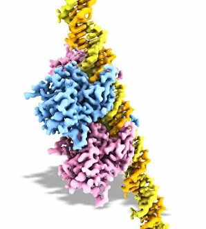
Background imageGenetic Collection: Tumour suppressor protein and DNA C017 / 3643

Tumour suppressor protein and DNA C017 / 3643
Tumour suppressor protein and DNA. Computer artwork showing a molecule of the tumour suppressor protein p53 (blue and pink) bound to a molecule of DNA (deoxyribonucleic acid, yellow and orange)

Background imageGenetic Collection: Homeodomain from Ubx and Exd protein C017 / 7006

Homeodomain from Ubx and Exd protein C017 / 7006
Structure of a DNA-bound Ultrabithorax (Ubx) and Extradenticle (Exd) homeodomain complex bound to DNA, showing how one of the helical regions fits into a major groove on the doulbe-helical DNA

Background imageGenetic Collection: Fertilisation, artwork C018 / 6998

Fertilisation, artwork C018 / 6998
Fertilisation. Computer artwork of numerous sperm cells (spermatozoa, white) surrounding an egg (round, centre). Only a single sperm cell can penetrate and fertilise the egg

Background imageGenetic Collection: Fertilisation, artwork C018 / 6995

Fertilisation, artwork C018 / 6995
Fertilisation. Computer artwork of a sperm cell (spermatozoon, purple) penetrating an egg (red, right)

Background imageGenetic Collection: Gigantism and dwarfism in farm animals C018 / 7060

Gigantism and dwarfism in farm animals C018 / 7060
Gigantism and dwarfism in farm animals. 1883 engraving depicting the occurrence of gigantism and dwarfism in farm animals

Background imageGenetic Collection: Sperm cell, artwork C018 / 6993

Sperm cell, artwork C018 / 6993
Sperm cell. Computer artwork of a sperm cell (spermatozoon), showing its torpedo-shaped head (left) and long tail that it uses for propulsion


All Professionally Made to Order for Quick Shipping

"Unlocking the Secrets: Exploring the Fascinating World of Genetics" In this captivating journey, we delve into the intricate realm of genetics, where computer screens display mesmerizing human genetic sequences. The double-stranded RNA molecule stands as a testament to the complex nature of our genetic makeup. Witness DNA transcription in action through a stunning molecular model, unraveling the process that shapes our very existence. Amidst this exploration, an elegant leopard in its melanistic phase rests gracefully on a log, reminding us of the diversity and beauty found within genes. Computer artwork showcases a beta DNA segment surrounded by spheres, symbolizing both innovation and interconnectedness within our genetic code. The nucleotide base matrix unveils patterns that hold profound significance in understanding hereditary traits. As we peer into abstract images of DNA molecules, we are reminded of their remarkable structure and infinite possibilities they hold for life itself. The intricacies continue with the visualization of nucleosome molecules – tiny structures that play a crucial role in organizing our genetic material. Amidst these wonders lies an HIV reverse transcription enzyme; it serves as a stark reminder of how they can shape not only life but also disease. Yet even amidst challenges, there is hope as scientists tirelessly work to decipher these complexities and find solutions. Ultimately, this captivating journey through various facets of genetics leaves us awestruck by its elegance and complexity. It reminds us that every living being carries within them an extraordinary story written in their DNA – an ancient language connecting all forms of life on Earth.