Fraud Blocker Skip to main content

Genetic Collection (page 6)

"Unlocking the Secrets: Exploring the Fascinating World of Genetics" In this captivating journey, we delve into the intricate realm of genetics

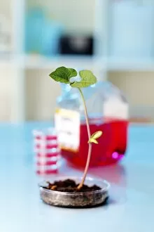
Background imageGenetic Collection: Genetic modification, conceptual image F008 / 2056

Genetic modification, conceptual image F008 / 2056
Genetically modified plant, conceptual image

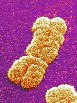
Background imageGenetic Collection: Human chromosome pair, SEM

Human chromosome pair, SEM
Human chromosome. Coloured scanning electron micrograph (SEM) of a human chromosome as a pair of identical copies called chromatids. These form as part of chromosome replication during cell division

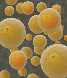
Background imageGenetic Collection: Genetically-modified pollen grains

Genetically-modified pollen grains
Genetically-modified pollen. Conceptual composite image of a coloured scanning electron micrograph (SEM) of maize pollen grains (Zea mays) labelled with biohazard symbols

Background imageGenetic Collection: DNA sequence, artwork F008 / 3293

DNA sequence, artwork F008 / 3293
DNA sequence, computer artwork

Background imageGenetic Collection: Genetics research, conceptual image F008 / 2093

Genetics research, conceptual image F008 / 2093
Genetics research, conceptual image

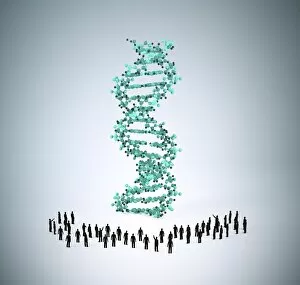
Background imageGenetic Collection: Human genome, conceptual artwork F008 / 3292

Human genome, conceptual artwork F008 / 3292
Human genome, conceptual computer artwork

Background imageGenetic Collection: Genetics research F008 / 3195

Genetics research F008 / 3195
Genetics research

Background imageGenetic Collection: Chromosomes, SEM

Chromosomes, SEM
Chromosomes. Coloured scanning electron micrograph (SEM) of two chromosomes. The SEM is overlaid on a DNA autoradiogram background

Background imageGenetic Collection: Giant chromosomes, light micrograph P657 / 0038

Giant chromosomes, light micrograph P657 / 0038
Giant chromosomes. Light micrograph of giant (polytene) chromosomes from a fruit fly (Drosophila busckii). These chromosomes are formed from repeated rounds of DNA (deoxyribonucleic acid)

Background imageGenetic Collection: Genetics research F008 / 3191

Genetics research F008 / 3191
Genetics research

Background imageGenetic Collection: Giant chromosomes, light micrograph P657 / 0037

Giant chromosomes, light micrograph P657 / 0037
Giant chromosomes. Light micrograph of giant (polytene) chromosomes from a fruit fly (Drosophila busckii). These chromosomes are formed from repeated rounds of DNA (deoxyribonucleic acid)

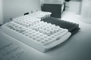
Background imageGenetic Collection: Microtubes of RNA samples F008 / 2041

Microtubes of RNA samples F008 / 2041
Microtubes of RNA samples

Background imageGenetic Collection: Human genome, conceptual artwork F008 / 3302

Human genome, conceptual artwork F008 / 3302
Human genome, conceptual computer artwork

Background imageGenetic Collection: Genetics research F008 / 3194

Genetics research F008 / 3194
Genetics research

Background imageGenetic Collection: Genetics research F005 / 0929

Genetics research F005 / 0929
MODEL RELEASED. Genetics research. Scientist performing gel electrophoresis under ultraviolet light
Background imageGenetic Collection: DNA nucleosome molecule

DNA nucleosome molecule
DNA nucleosome, molecular model. This is the fundamental repeating unit used to package DNA (deoxyribonucleic acid) inside cell nuclei

Background imageGenetic Collection: Genetics research F005 / 0930

Genetics research F005 / 0930
MODEL RELEASED. Genetics research. Scientist performing gel electrophoresis under ultraviolet light

Background imageGenetic Collection: Medical test tube with blood, artwork

Medical test tube with blood, artwork
Computer artwork of a medical test tube with blood samples and a DNA helix, depicting dna profiling, genetic testing and other DNA based tests

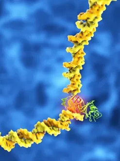
Background imageGenetic Collection: TATA box-binding protein complex C014 / 0867

TATA box-binding protein complex C014 / 0867
TATA box-binding protein complex. Molecular model showing a TATA box-binding protein (TBP, khaki) complexed with a strand of DNA (deoxyribonucleic acid)

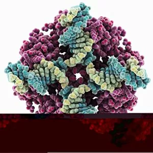
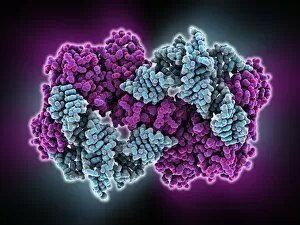
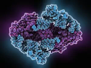
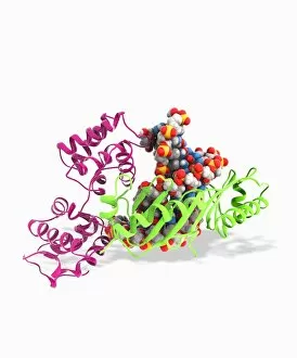
Background imageGenetic Collection: E. coli Holliday junction complex C014 / 0878

E. coli Holliday junction complex C014 / 0878
E. coli Holliday junction complex. Molecular model of a RuvA protein (dark pink) in complex with a Holliday junction between homologous strands of DNA (deoxyribonucleic acid)

Background imageGenetic Collection: Genetic research, conceptual image C014 / 1256

Genetic research, conceptual image C014 / 1256
Genetic research. Conceptual image of a molecular model of a strand of DNA (deoxyribonucleic acid) being held on a human hand

Background imageGenetic Collection: DNA supercoil, artwork

DNA supercoil, artwork
DNA supercoils. Computer artwork showing a supercoiled strand of DNA (deoxyribonucleic acid). Supercoiling is important in a number of biological processes

Background imageGenetic Collection: Tyrosyl-tRNA synthetase molecule

Tyrosyl-tRNA synthetase molecule
Tyrosyl-tRNA synthetase protein molecule. Molecular model showing bacterial tyrosyl-tRNA synthetase complexed with tyrosyl tRNA (transfer ribonucleic acid)

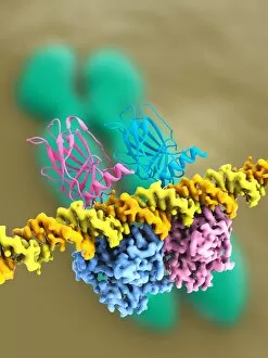
Background imageGenetic Collection: Tumour suppressor protein and DNA C017 / 3645

Tumour suppressor protein and DNA C017 / 3645
Tumour suppressor protein and DNA. Computer artwork showing a molecule of the tumour suppressor protein p53 (blue and pink) bound to a molecule of DNA (deoxyribonucleic acid, yellow and orange)

Background imageGenetic Collection: Synthetic biology, conceptual artwork C018 / 0907

Synthetic biology, conceptual artwork C018 / 0907
Synthetic biology, conceptual illustration. Lab set-up inside a yeast cell

Background imageGenetic Collection: DNA 6-way junction, artwork C014 / 2587

DNA 6-way junction, artwork C014 / 2587
DNA 6-way junction. Computer artwork of a synthetic assemblage of nucleic acids which are useful in the design of nanostructures

Background imageGenetic Collection: Tryptophanyl-tRNA synthetase molecule

Tryptophanyl-tRNA synthetase molecule
Tryptophanyl-tRNA synthetase protein molecule. Molecular model showing human tryptophanyl-tRNA synthetase complexed with tryptophan tRNA (transfer ribonucleic acid)

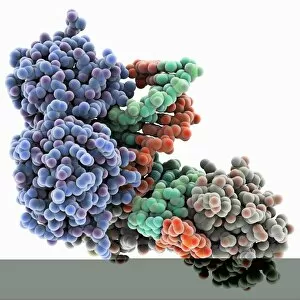
Background imageGenetic Collection: Pho4 transcription factor bound to DNA C014 / 0861

Pho4 transcription factor bound to DNA C014 / 0861
Pho4 transcription factor bound to DNA. Molecular model showing phosphate system positive regulatory protein (Pho4) (blue and green) bound to a strand of DNA (deoxyribonucleic acid, red and purple)

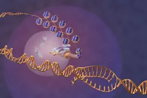
Background imageGenetic Collection: Gene expression, artwork

Gene expression, artwork
Gene expression. Computer artwork showing the process of transcription, the first stage or gene expression. Here, a chromosome (distance)

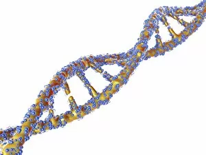
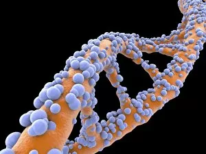
Background imageGenetic Collection: DNA molecule, artwork F007 / 1996

DNA molecule, artwork F007 / 1996
DNA molecule, computer artwork

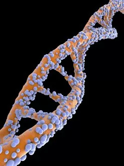
Background imageGenetic Collection: DNA molecule, artwork F007 / 1994

DNA molecule, artwork F007 / 1994
DNA molecule, computer artwork

Background imageGenetic Collection: DNA molecule, artwork F007 / 1995

DNA molecule, artwork F007 / 1995
DNA molecule, computer artwork

Background imageGenetic Collection: DNA molecule, artwork F007 / 1991

DNA molecule, artwork F007 / 1991
DNA molecule, computer artwork

Background imageGenetic Collection: DNA molecule, artwork F007 / 1992

DNA molecule, artwork F007 / 1992
DNA molecule, computer artwork

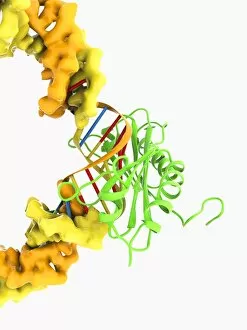
Background imageGenetic Collection: TATA box-binding protein complex C017 / 7090

TATA box-binding protein complex C017 / 7090
TATA box-binding protein complex. Molecular model showing a TATA box-binding protein (TBP, green) complexed with a strand of DNA (deoxyribonucleic acid, spheres) and transcription factor IIB

Background imageGenetic Collection: TATA box-binding protein complex C017 / 7085

TATA box-binding protein complex C017 / 7085
TATA box-binding protein complex. Molecular model showing a TATA box-binding protein (TBP, green) complexed with a strand of DNA (deoxyribonucleic acid, yellow) and transcription factor IIB

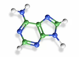
Background imageGenetic Collection: Adenine molecule, artwork C017 / 7199

Adenine molecule, artwork C017 / 7199
Adenine molecule. Computer artwork showing the structure of a molecule of the nucleobase adenine. Atoms are colour-coded spheres: carbon (green), nitrogen (blue), and oxygen (white)

Background imageGenetic Collection: Junk DNA, conceptual image

Junk DNA, conceptual image. Computer artwork of damaged DNA (deoxyribonucleic acid) in a rubbish bin. DNA contains sections called genes that encode the bodys genetic information

Background imageGenetic Collection: Genetic code, artwork F006 / 8998

Genetic code, artwork F006 / 8998
Genetic code, computer artwork

Background imageGenetic Collection: Genetic code, artwork F006 / 8997

Genetic code, artwork F006 / 8997
Genetic code, computer artwork

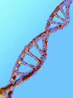
Background imageGenetic Collection: DNA molecule, artwork F006 / 8969

DNA molecule, artwork F006 / 8969
DNA molecule, computer artwork

Background imageGenetic Collection: DNA molecules, artwork F007 / 0044

DNA molecules, artwork F007 / 0044
DNA molecules, computer artwork

Background imageGenetic Collection: DNA molecules, artwork F007 / 0045

DNA molecules, artwork F007 / 0045
DNA molecules, computer artwork

Background imageGenetic Collection: TATA box-binding protein complex C017 / 7083

TATA box-binding protein complex C017 / 7083
TATA box-binding protein complex. Molecular model showing a TATA box-binding protein (TBP, green) complexed with a strand of DNA (deoxyribonucleic acid, yellow) and transcription factor IIB

Background imageGenetic Collection: DNA molecules, artwork F006 / 8775

DNA molecules, artwork F006 / 8775
DNA molecules, computer artwork

Background imageGenetic Collection: DNA molecules, artwork F006 / 8776

DNA molecules, artwork F006 / 8776
DNA molecules, computer artwork


All Professionally Made to Order for Quick Shipping

"Unlocking the Secrets: Exploring the Fascinating World of Genetics" In this captivating journey, we delve into the intricate realm of genetics, where computer screens display mesmerizing human genetic sequences. The double-stranded RNA molecule stands as a testament to the complex nature of our genetic makeup. Witness DNA transcription in action through a stunning molecular model, unraveling the process that shapes our very existence. Amidst this exploration, an elegant leopard in its melanistic phase rests gracefully on a log, reminding us of the diversity and beauty found within genes. Computer artwork showcases a beta DNA segment surrounded by spheres, symbolizing both innovation and interconnectedness within our genetic code. The nucleotide base matrix unveils patterns that hold profound significance in understanding hereditary traits. As we peer into abstract images of DNA molecules, we are reminded of their remarkable structure and infinite possibilities they hold for life itself. The intricacies continue with the visualization of nucleosome molecules – tiny structures that play a crucial role in organizing our genetic material. Amidst these wonders lies an HIV reverse transcription enzyme; it serves as a stark reminder of how they can shape not only life but also disease. Yet even amidst challenges, there is hope as scientists tirelessly work to decipher these complexities and find solutions. Ultimately, this captivating journey through various facets of genetics leaves us awestruck by its elegance and complexity. It reminds us that every living being carries within them an extraordinary story written in their DNA – an ancient language connecting all forms of life on Earth.