Fraud Blocker Skip to main content

Gametes Collection

Gametes, the tiny yet powerful reproductive cells, play a crucial role in the cycle of life

Background imageGametes Collection: Geranium anther, SEM

Geranium anther, SEM
Geranium anther. Coloured scanning electron micrograph (SEM) of pollen (pink) on the anther of a geranium flower (Geranium sp.)

Background imageGametes Collection: Honeybee leg, SEM

Honeybee leg, SEM
Honey bee leg. Coloured scanning electron micrograph (SEM) of the leg of a honeybee (Apis mellifera) covered in pollen grains (round). The dense coating of hairs has trapped the pollen

Background imageGametes Collection: Geranium pollen, SEM

Geranium pollen, SEM
Geranium pollen. Coloured scanning electron micrograph (SEM) of pollen (pink) on the stamen of a geranium flower (Geranium sp.)

Background imageGametes Collection: IUD contraceptive and sperm cells

IUD contraceptive and sperm cells. Computer artwork of an intrauterine device (IUD, red and white, centre) in a womans uterus, surrounded by sperm cells (white, not to scale)

Background imageGametes Collection: Picture No. 11675582

Picture No. 11675582
Scanning Electron micrograph (SEM): Lily Pollen grains. Date:

Background imageGametes Collection: Pollen on a bee leg, SEM

Pollen on a bee leg, SEM
Pollen on a bee leg. Coloured scanning electron micrograph (SEM) of pollen grains (pale purple) on the leg of a bee (superfamily Apoidea). Pollen grains are the male sex cells of a flowering plant

Background imageGametes Collection: Philadelphia fleabane pollen grains, SEM

Philadelphia fleabane pollen grains, SEM
Philadelphia fleabane (Erigeron philadelphicus) pollen grains, coloured scanning electron micrograph (SEM). Pollen grains are reproductive structures produced by the male parts of flowering plants

Background imageGametes Collection: Gorse stigma with pollen grains, SEM

Gorse stigma with pollen grains, SEM
Gorse (Ulex europaeus) stigma (green) with pollen grains (yellow), coloured scanning electron micrograph (SEM). Pollen grains are reproductive structures produced by the male parts of flowering

Background imageGametes Collection: Forsythia pollen grains, SEM

Forsythia pollen grains, SEM
Forsythia pollen grains arranged in a spiral, coloured scanning electron micrograph (SEM). Pollen grains are reproductive structures produced by the male parts of flowering plants

Background imageGametes Collection: Chickweed pollen grains, SEM

Chickweed pollen grains, SEM
Chickweed pollen grains. Coloured scanning electron micrograph (SEM) of pollen grains (green) from a chickweed flower (Stellaria media)

Background imageGametes Collection: Flame lily pollen, SEM

Flame lily pollen, SEM
Flame lily (Gloriosa sp.) pollen grains, coloured scanning electron micrograph (SEM). The grains have a highly sculptured outer wall (exine)

Background imageGametes Collection: Hellebore pollen, SEM

Hellebore pollen, SEM
Hellebore (Helleborus sp.) pollen grains, coloured scanning electron micrograph (SEM). Magnification: x2000 when printed at 10 centimetres wide

Background imageGametes Collection: Germinating lily pollen, SEM

Germinating lily pollen, SEM
Germinating lily pollen. Coloured scanning electron micrograph (SEM) of lily (Lilium sp.) pollen grains that have sprouted pollen tubes. Pollen contains the male sex cells of a plant

Background imageGametes Collection: Pollination, SEM

Pollination, SEM
Pollination. Coloured scanning electron micrograph (SEM) of part of a flowers pistil with pollen grains attached. The pistil is the female reproductive part of a flower

Background imageGametes Collection: Marrow and forget-me-not pollen, SEM

Marrow and forget-me-not pollen, SEM
Pollen grains. Coloured scanning electron micrograph (SEM) of a pollen grain from a marrow plant (Cucurbita sp. left) and a forget-me-not plant (Myosotis sp. lower right)

Background imageGametes Collection: Hollyhock pollen grain, SEM

Hollyhock pollen grain, SEM
Hollyhock pollen grain. Coloured scanning electron micrograph (SEM) of a pollen grain from a hibiscus (Alcea setosa) flower. Pollen grains are the male sex cells of a flowering plant

Background imageGametes Collection: Verbena pollen, SEM

Verbena pollen, SEM
Verbena pollen. Coloured scanning electron micrograph (SEM) of pollen grains from verbena bonariensis. Verbena bonariensis is a tall, slender-stemmed perennial

Background imageGametes Collection: Convolvulus pollen grains, SEM

Convolvulus pollen grains, SEM
Convolvulus pollen grains. Coloured scanning electron micrograph (SEM) of pollen grains from a convolvulus flower. Convolvulus is a genus of about 200 to 250 species of flowering plants in

Background imageGametes Collection: Water lily pollen grains, SEM

Water lily pollen grains, SEM
Water lily pollen grains. Coloured scanning electron micrograph (SEM) of pollen grains from a water lily flower. Nymphaeaceae is a family of flowering plants, commonly called water lilies

Background imageGametes Collection: Human sperm cells

Human sperm cells
Sperm. Computer artwork of human sperm (spermatozoa), the sex cells of the male. Each sperm is about 0.065 millimetres in length

Background imageGametes Collection: Human genetic engineering

Human genetic engineering. Conceptual computer artwork representing the genetic engineering of humans. Sperm cells labelled with bar codes are seen

Background imageGametes Collection: Carnation flower pistil, SEM C016 / 9762

Carnation flower pistil, SEM C016 / 9762
Carnation (Dianthus sp.) pistil, coloured scanning electron micrograph (SEM). Pistils hold a plants female reproductive structures

Background imageGametes Collection: Carnation flower pistil, SEM C016 / 9759

Carnation flower pistil, SEM C016 / 9759
Carnation (Dianthus sp.) pistil, coloured scanning electron micrograph (SEM). Pistils hold a plants female reproductive structures

Background imageGametes Collection: Carnation flower pistil, SEM C016 / 9760

Carnation flower pistil, SEM C016 / 9760
Carnation (Dianthus sp.) pistil, coloured scanning electron micrograph (SEM). Pistils hold a plants female reproductive structures

Background imageGametes Collection: Sperm, SEM

Sperm, SEM
Sperm. Coloured scanning electron micrograph (SEM) of human sperm (spermatozoa), the sex cells of the male. Sperm are about 0.065 millimetres in length

Background imageGametes Collection: Hibiscus flower pistil, SEM C016 / 9758

Hibiscus flower pistil, SEM C016 / 9758
Hibiscus flower pistil. Coloured scanning electron micrograph (SEM) of a Hibiscus sp. flower pistil. Pistils hold a plants female reproductive structures

Background imageGametes Collection: Fungal spores on pollen grain, SEM

Fungal spores on pollen grain, SEM
Fungal spores on pollen grain. Coloured scanning electron micrograph (SEM) showing fungal spores (round) on a pollen grain from a goji berry (wolfberry, Lycium sp.) plant

Background imageGametes Collection: Carnation flower pistil, SEM C016 / 9763

Carnation flower pistil, SEM C016 / 9763
Carnation (Dianthus sp.) pistil, coloured scanning electron micrograph (SEM). Pistils hold a plants female reproductive structures

Background imageGametes Collection: Hibiscus flower pistil, SEM C016 / 9757

Hibiscus flower pistil, SEM C016 / 9757
Hibiscus flower pistil. Coloured scanning electron micrograph (SEM) of a Hibiscus sp. flower pistil. Pistils hold a plants female reproductive structures

Background imageGametes Collection: Carnation flower pistil, SEM C016 / 9761

Carnation flower pistil, SEM C016 / 9761
Carnation (Dianthus sp.) pistil, coloured scanning electron micrograph (SEM). Pistils hold a plants female reproductive structures

Background imageGametes Collection: Fertilisation, artwork C016 / 7516

Fertilisation, artwork C016 / 7516
Fertilisation. Cutaway computer artwork of a sperm cell (spermatozoa) penetrating an egg (large). The nucleus of the sperm cell will fuse with the egg and its DNA (deoxyribonucleic acid)

Background imageGametes Collection: Ovulation, artwork C016 / 7537

Ovulation, artwork C016 / 7537
Ovulation, computer artwork

Background imageGametes Collection: Cowslip pollen grain, SEM C016 / 9031

Cowslip pollen grain, SEM C016 / 9031
Cowslip pollen grain. Coloured scanning electron micrograph (SEM) of pollen grain from a common cowslip (Primula veris) flower. Pollen grains are the male sex cells of a flowering plant

Background imageGametes Collection: Cowslip pollen grain, SEM C016 / 9030

Cowslip pollen grain, SEM C016 / 9030
Cowslip pollen grain. Coloured scanning electron micrograph (SEM) of pollen grain from a common cowslip (Primula veris) flower. Pollen grains are the male sex cells of a flowering plant

Background imageGametes Collection: Dwarf checkermallow pollen grain, SEM C016 / 9438

Dwarf checkermallow pollen grain, SEM C016 / 9438
Dwarf checkermallow pollen grain. Coloured scanning electron micrograph (SEM) of a pollen grain from a dwarf checkermallow (Sidalcea malviflora) flower

Background imageGametes Collection: Fertilisation, artwork C016 / 7515

Fertilisation, artwork C016 / 7515
Fertilisation. Computer artwork of a sperm cell (spermatozoa) penetrating an egg (large)

Background imageGametes Collection: Carnation flower pistil, SEM C016 / 0127

Carnation flower pistil, SEM C016 / 0127
Carnation (Dianthus sp.) pistil, coloured scanning electron micrograph (SEM). Pistils hold a plants female reproductive structures

Background imageGametes Collection: Carnation flower pistil, SEM C016 / 0131

Carnation flower pistil, SEM C016 / 0131
Carnation (Dianthus sp.) pistil, coloured scanning electron micrograph (SEM). Pistils hold a plants female reproductive structures

Background imageGametes Collection: Camellia pollen, SEM C015 / 9103

Camellia pollen, SEM C015 / 9103
Camellia pollen grains. Coloured scanning electron micrograph (SEM) of pollen grains from a Camellia sp. flower. The grains have a highly sculpted outer wall (exine)

Background imageGametes Collection: Carnation flower pistil, SEM C016 / 0119

Carnation flower pistil, SEM C016 / 0119
Carnation (Dianthus sp.) pistil, coloured scanning electron micrograph (SEM). Pistils hold a plants female reproductive structures

Background imageGametes Collection: Flower pistils, SEM C016 / 0053

Flower pistils, SEM C016 / 0053
Flower pistils, coloured scanning electron micrograph (SEM). Pistils hold a plants female reproductive structures. They contain one or more carpels, which, in turn, contain a stigma

Background imageGametes Collection: Rose pistil, SEM C015 / 9173

Rose pistil, SEM C015 / 9173
Rose pistil. Coloured scanning electron micrograph (SEM) of a rose (Rosa sp.) flower pistil. Pistils hold a plants female reproductive structures

Background imageGametes Collection: Rose pistil, SEM C015 / 9181

Rose pistil, SEM C015 / 9181
Rose pistil. Coloured scanning electron micrograph (SEM) of a rose (Rosa sp.) flower pistil. Pistils hold a plants female reproductive structures

Background imageGametes Collection: Geranium pollen, SEM C015 / 9155

Geranium pollen, SEM C015 / 9155
Geranium pollen. Coloured scanning electron micrograph (SEM) of pollen (pink) on the stamen of a geranium flower (Geranium sp.)

Background imageGametes Collection: Camellia pollen, SEM C015 / 9101

Camellia pollen, SEM C015 / 9101
Camellia pollen grains. Coloured scanning electron micrograph (SEM) of pollen grains from a Camellia sp. flower. The grains have a highly sculpted outer wall (exine)

Background imageGametes Collection: Camellia pollen, SEM C015 / 9089

Camellia pollen, SEM C015 / 9089
Camellia pollen grains. Coloured scanning electron micrograph (SEM) of pollen grains from a Camellia sp. flower. The grains have a highly sculpted outer wall (exine)


All Professionally Made to Order for Quick Shipping

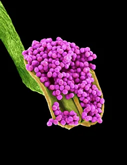
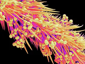
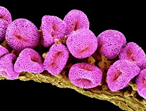
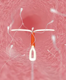
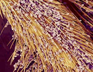
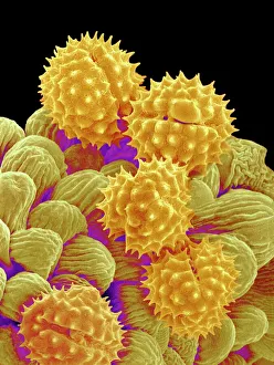
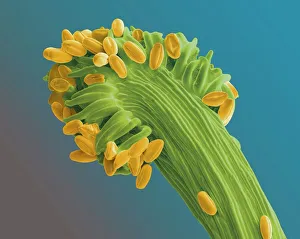
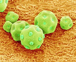
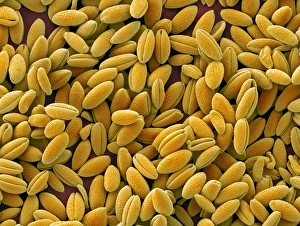
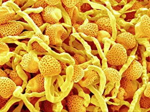
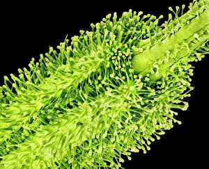
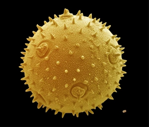
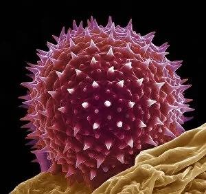
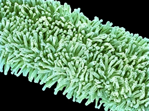
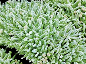
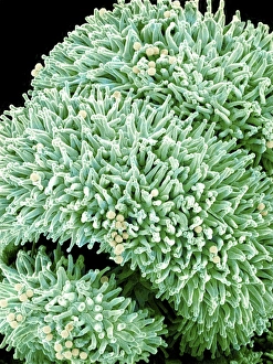
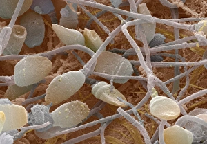
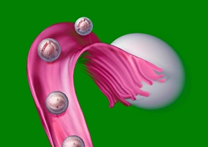
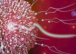
Gametes, the tiny yet powerful reproductive cells, play a crucial role in the cycle of life. Captured under the lens of a scanning electron microscope (SEM), these microscopic wonders reveal their intricate beauty and significance. In one captivating image, we observe the delicate structure of a Geranium anther. Its vibrant colors and fine details showcase nature's artistry at its finest. This anther holds within it countless Geranium pollen grains, each carrying the potential to create new life. Moving on to another SEM masterpiece, we witness a Honeybee leg adorned with pollen grains collected during its pollination journey. These minuscule particles cling to every hair-like structure on the bee's leg, showcasing how these industrious insects inadvertently aid in plant reproduction while seeking nectar. Zooming closer into nature's marvels, Philadelphia fleabane pollen grains come into focus through SEM imagery. Their unique shapes and textures hint at their adaptability for efficient dispersal by wind or animal vectors – ensuring successful fertilization across vast distances. Gorse stigma with pollen grains presents us with yet another mesmerizing sight under SEM observation. The interplay between this flower's receptive surface and meticulously crafted pollen grains showcases evolution's ingenuity in facilitating cross-pollination among plants. Shifting gears from flora to contraception methods brings us face-to-face with IUD contraceptive and sperm cells' juxtaposition. This thought-provoking image serves as a reminder that gametes not only hold promise for creation but also highlight humanity's quest for reproductive control. As our exploration continues through SEM imagery, Forsythia pollen grains captivate us with their elegance and symmetry. Each grain acts as a messenger transporting genetic material from one flower to another – perpetuating biodiversity in our natural world. Chickweed pollen grains make their appearance next – small yet significant contributors to plant reproduction through insect-mediated pollination processes. Their presence reminds us of the interconnectedness between various organisms within ecosystems.