
Fungi Collection (#46)
"Fungi: The Fascinating World of Mushroom Kingdom" Delicate and mysterious, mushrooms captivate our imagination with their intricate forms and colors


All Professionally Made to Order for Quick Shipping
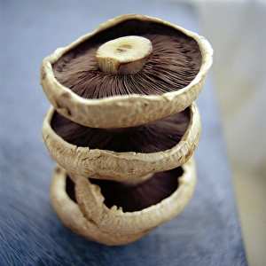
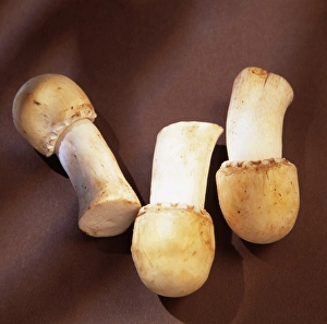
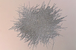
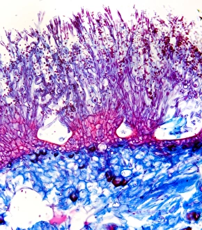
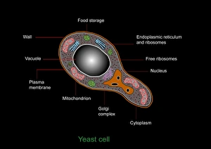
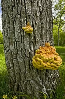
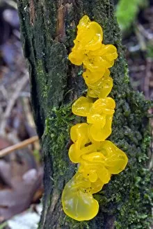
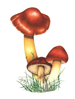
"Fungi: The Fascinating World of Mushroom Kingdom" Delicate and mysterious, mushrooms captivate our imagination with their intricate forms and colors. Zooming in on mushroom spores reveals a mesmerizing close-up view that resembles a miniature galaxy. Witness the birth of life as a budding yeast cell emerges, ready to multiply and thrive. Step back in time with a vintage photograph showcasing the astonishing diversity of mushrooms from 1913. Enter the enchanting realm of fly agaric mushrooms, known for their vibrant red caps speckled with white dots. Explore the hidden world of penicillin fungus through an electron microscope's lens - nature's own antibiotic factory. Behold the captivating beauty of Aspergillus nidulans fungus thriving in its cultured environment, resembling an otherworldly landscape. Discover the culinary delight that is Cep mushroom (Boletus edulis), cherished by chefs for its rich flavor and meaty texture. Mushrooms come in all shapes and sizes, each one boasting unique characteristics that make them truly extraordinary. Candida fungus takes center stage under scanning electron microscopy, revealing its intricate structure like never before seen by human eyes. Coral Spot Fungus (Nectria cinnabarina) fruiting bodies emerge on a Sycamore twig in Powys, Wales – nature's artistry at work. Explore the delicate intricacy of mushroom gills under high-resolution SEM imaging – nature's design perfected over millions of years.