Fraud Blocker Skip to main content

Follicles Collection

"Fascinating Follicles: Exploring the Wonders of Hair and Reproduction" Picture No

Background imageFollicles Collection: Picture No. 10876997

Picture No. 10876997
Scanning Electron Micrograph (SEM): Human Skin with Hair Follicles Date:

Background imageFollicles Collection: Sycosis

Sycosis
5855392 Sycosis; (add.info.: Sycosis barbae and Sycosis capilli, chronic inflammation of the hair follicles, especially of the beard and head)

Background imageFollicles Collection: The Skin (colour litho)

The Skin (colour litho)
2799331 The Skin (colour litho) by English School, (19th century); Private Collection; (add.info.: The Skin. Illustration for The Home Handbook of Domestic Hygiene)

Background imageFollicles Collection: Diagram showing the interaction between female sexual organs and the brain, on one side

Diagram showing the interaction between female sexual organs and the brain, on one side, the normal reproductive cycle, and on the other, the effect of the contraceptive pill

Background imageFollicles Collection: Diagram illustrating the two layers of human skin, epidermis, dermis and hair follicle

Diagram illustrating the two layers of human skin, epidermis, dermis and hair follicle

Background imageFollicles Collection: Eyelash hairs, SEM

Eyelash hairs, SEM
Eyelash hairs and skin. Coloured scanning electron micrograph (SEM) of eyelash hairs growing from the surface of human skin

Background imageFollicles Collection: Human and animal hair and follicles magnified

Human and animal hair and follicles magnified.. Hair and follicles magnified- Human 1, insect and quadruped 2, and roe deer 3

Background imageFollicles Collection: Scalp skin showing hair follicles, LM

Scalp skin showing hair follicles, LM
Scalp skin. Light micrograph of a section through skin from the scalp. Numerous hair shafts are seen (purple rings), which are surrounded by connective tissue (dark blue)

Background imageFollicles Collection: Hairy scalp skin, light micrograph

Hairy scalp skin, light micrograph
Hairy scalp skin. Light micrograph of a section through healthy skin from a human scalp, showing the numerous hair follicles. The scalp is an example of hairy skin

Background imageFollicles Collection: Alopecia, artwork C016 / 7523

Alopecia, artwork C016 / 7523
Alopecia. Computer artwork showing hair being lost in a person with alopecia

Background imageFollicles Collection: Primordial egg follicles in an ovary

Primordial egg follicles in an ovary
Light micrograph of three primordial follicles (at centre) surrounded by flattened stroma cells in the human ovary. These follicles will form into oocytes which become eggs

Background imageFollicles Collection: Thyroid follicles, light micrograph

Thyroid follicles, light micrograph
Thyroid follicles. Light micrograph of a section through a thyroid gland showing the follicles. The follicles are lined by a single layer of cuboidal epithelial cells

Background imageFollicles Collection: Appendix, light micrograph C016 / 0510

Appendix, light micrograph C016 / 0510
Appendix. Light micrograph of a section through a human appendix. The appendix is a blind-ending small diameter tubular extension of the gut, arising from the caecum of the large bowel

Background imageFollicles Collection: Ovarian primordial follicles, micrograph C016 / 0518

Ovarian primordial follicles, micrograph C016 / 0518
Ovarian primordial follicles. Light micrograph of a section through the cortex of a neonatal ovary, showing clusters of primordial follicles each containing a single oocyte (egg cell)

Background imageFollicles Collection: Thyroid gland, SEM C015 / 6419

Thyroid gland, SEM C015 / 6419
Thyroid gland. Coloured scanning electron micrograph (SEM) of a fracture through the thyroid gland revealing several follicles (green and blue). Between the follicles is connective tissue (pink)

Background imageFollicles Collection: Mouse tail, light micrograph

Mouse tail, light micrograph
Mouse tail. Light micrograph of a cross-section through a mouses tail. At centre is the tail vein, which contains red blood cells. A smaller vein (purple squiggle) is seen at top centre left

Background imageFollicles Collection: Human skin, polarised light micrograph

Human skin, polarised light micrograph
Human skin. Polarised light micrograph of a section through human skin showing hair follicles (black). The top layer of the skin

Background imageFollicles Collection: Eyebrow hair, SEM

Eyebrow hair, SEM
Eyebrow hair and skin. Coloured scanning electron micrograph (SEM) of eyebrow hair growing from the surface of human skin

Background imageFollicles Collection: Hair follicles, SEM

Hair follicles, SEM
Hair follicles. Coloured scanning electron micrograph (SEM) of a section through freeze- fractured hair follicles in the skin

Background imageFollicles Collection: Stubble hair, SEM

Stubble hair, SEM
Stubble hair and skin. Coloured scanning electron micrograph (SEM) of stubble hair growing from the surface of human skin

Background imageFollicles Collection: Thyroid gland, light micrograph

Thyroid gland, light micrograph
Thyroid gland. Coloured light micrograph of a thyroid gland showing the follicles. The follicles are lined by a single layer of cuboidal epithelial cells (purple, dark nuclei)

Background imageFollicles Collection: Thyroid follicles

Thyroid follicles. Computer artwork of cells of thyroid follicles (pink, purple) superimposed on an artwork of the thyroid gland (red, centre)

Background imageFollicles Collection: Follicles in a human ovary, micrograph

Follicles in a human ovary, micrograph
Developing follicles in a human ovary, light micrograph. Three developing Graafian follicles are seen. A Graafian follicle is a structure in the ovaries from which an ovum (egg cell) is released

Background imageFollicles Collection: Artwork of ovum (egg) development in womans ovary

Artwork of ovum (egg) development in womans ovary
Ovum development. Illustration showing the development of a human ovum (egg) in an ovary (pink, at lower centre) during the menstrual cycle

Background imageFollicles Collection: Graafian follicles, light micrograph

Graafian follicles, light micrograph
Developing Graafian follicles. Light micrograph of a sectioned human ovary, showing two developing Graafian follicles. A Graafian follicle is a structure in the ovaries from which an ovum (egg cell)

Background imageFollicles Collection: Palm

Palm. Magnified section of palm, historical artwork. The external ridged surface is the cuticle, with the thin layer of skin below this known as the rete mucosum, or epidermis

Background imageFollicles Collection: Magnified section of scalp

Magnified section of scalp, historical artwork. The external surface is the cuticle, with the layer of skin below this known as the rete mucosum, or epidermis

Background imageFollicles Collection: Thyroid follicles, artwork

Thyroid follicles, artwork
Thyroid gland. Computer artwork of a thyroid gland (red, centre), superimposed on an image of cells of thyroid follicles (green, blue)

Background imageFollicles Collection: Upper lip skin, light micrograph

Upper lip skin, light micrograph. Numerous small hair follicles (purple) can be seen. Magnification: x60 when printed at 10 centimetres wide

Background imageFollicles Collection: Hair follicles, light micrograph

Hair follicles, light micrograph. The hair shaft (orange) is surrounded by an internal root sheath (yellow), an external root sheath (pink) and connective tissue (brown)

Background imageFollicles Collection: Thyroid, light micrograph

Thyroid, light micrograph
Thyroid. Light micrograph of a section through a thyroid gland. The dark purple areas are follicles that are filled with a storage form form of the thyroid hormones

Background imageFollicles Collection: Cat lymph gland, light micrograph

Cat lymph gland, light micrograph

Background imageFollicles Collection: Lymph gland, light micrograph

Lymph gland, light micrograph

Background imageFollicles Collection: Inflamed lymph gland, light micrograph

Inflamed lymph gland, light micrograph

Background imageFollicles Collection: Mammalian ovary, light micrograph

Mammalian ovary, light micrograph
Mammalian ovary. Light micrograph of a transverse section through a mammalian ovary, the reproductive structure that produces the female sex cells (ova)

Background imageFollicles Collection: Blocked pore in acne, artwork

Blocked pore in acne, artwork
Blocked pore in acne, computer artwork. Sweat pore (centre) that has become blocked (centre) due to acne. Acne is a skin disorder in which the sebaceous glands of the skin become inflamed (red areas)

Background imageFollicles Collection: Bacterial skin infections, artwork

Bacterial skin infections, artwork
Bacterial skin infections, computer artwork. Round (green, cocci) and cylindrical (blue, bacilli) bacteria on skin, with hair shafts also seen

Background imageFollicles Collection: Graafian follicle, light micrograph

Graafian follicle, light micrograph
Graafian follicle. Light micrograph of a section through a mature Graafian follicle from a mammalian ovary. This contains the secondary oocyte (round object at centre)

Background imageFollicles Collection: Microscopic Objects

Microscopic Objects
A variety of living and non- living objects magnified through a microscope


All Professionally Made to Order for Quick Shipping

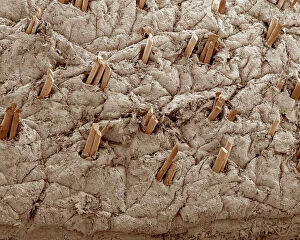
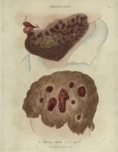
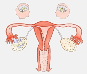
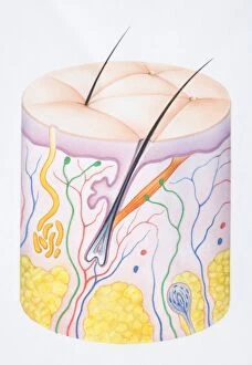
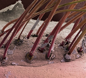
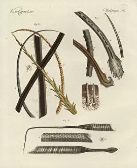
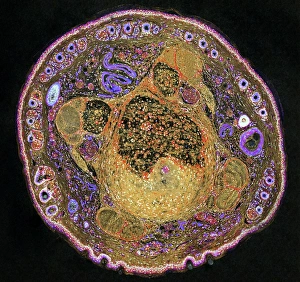
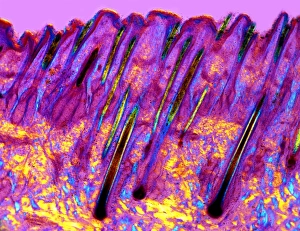
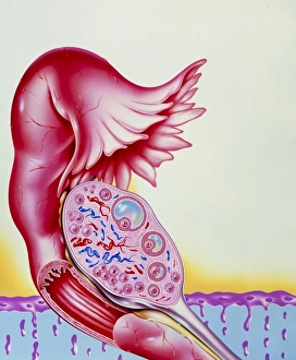
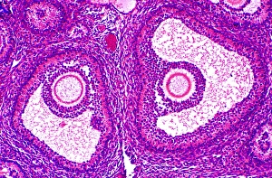
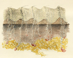
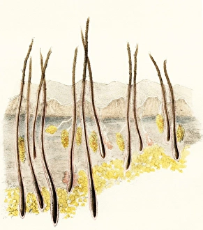
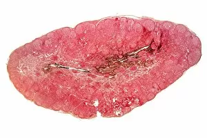
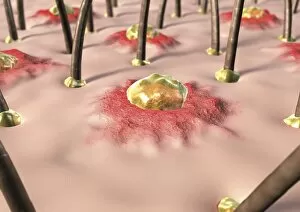
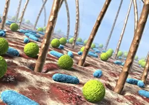
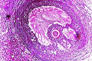
"Fascinating Follicles: Exploring the Wonders of Hair and Reproduction" Picture No. 10876997: Unveiling the intricate world of follicles, where secrets lie beneath the surface. The Skin (colour litho): Delving into the layers of human skin, discovering the hidden realms of epidermis, dermis, and hair follicles. Human and animal hair and follicles magnified: Zooming in on nature's incredible diversity, witnessing the microscopic wonders that adorn our bodies. Diagram showing the interaction between female sexual organs and the brain: Unraveling the complex dance between hormones and reproductive cycles, a delicate balance orchestrated by our bodies. The effect of contraceptive pills on reproductive cycles: Shedding light on how modern medicine alters nature's course, providing women with choices while understanding their impact on fertility. Eyelash hairs (SEM): Peering through a scanning electron microscope to witness eyelashes up close – tiny strands that frame our eyes with elegance. Scalp skin showing hair follicles (LM): Journeying into our scalps to explore where luscious locks originate from – an intricate network of nourishing follicles working tirelessly to grow healthy hair. Ovarian follicles (light micrograph): Glimpsing inside ovaries to marvel at these precious structures housing potential life within them – a testament to female fertility. Female reproductive system artwork: Celebrating womanhood through artistic depictions of this awe-inspiring system that allows for creation itself - a masterpiece crafted by evolution. Female reproductive system artwork x3 : Emphasizing its significance threefold; appreciating its complexity as it harmoniously connects various organs in pursuit of new beginnings. Intriguingly interconnected yet uniquely diverse, "follicles" unveil captivating stories about both beauty and reproduction within us.