Fraud Blocker Skip to main content

Fixation Collection

"Fixation: Restoring Strength and Stability through Precision" In the realm of medicine

Background imageFixation Collection: Picture No. 10899510

Picture No. 10899510
Lucerne / alfalfa / alphalpha (Medicago sativa ssp. sativa) Date:

Background imageFixation Collection: Pinned broken arm, X-ray C017 / 7869

Pinned broken arm, X-ray C017 / 7869
Pinned broken arm. X-ray of the arm of a patient that has been internally fixed after fracturing both of the lower arm bones, the ulna and radius

Background imageFixation Collection: Fixed arm fracture, X-ray C017 / 7265

Fixed arm fracture, X-ray C017 / 7265
Fixed arm fracture. X-ray of the elbow of a 4 year old female that has had a displaced supracondylar fracture fixed. The fracture is at the base of the humerus, the upper arm bone (upper frame)

Background imageFixation Collection: Kirschner wires, artwork C016 / 6994

Kirschner wires, artwork C016 / 6994
Kirschner wires. Artwork of a hand holding Kirschner wires, also known as K-wires. The bones of the hand and wrist are also shown

Background imageFixation Collection: Scoliosis treatment, X-ray C017 / 7156

Scoliosis treatment, X-ray C017 / 7156
Scoliosis treatment. X-ray of the spine of a 46 year old female patient that has been fixed to treat scoliosis (sideways curvature)

Background imageFixation Collection: Pinned jaw fracture, X-ray C017 / 7982

Pinned jaw fracture, X-ray C017 / 7982
Pinned jaw fracture. X-ray of the head of a 22 year old male patient who has had a fractured mandible (lower jaw bone) pinned (lower left)

Background imageFixation Collection: Pinned broken ankle, X-ray C017 / 7978

Pinned broken ankle, X-ray C017 / 7978
Pinned broken ankle. X-ray of the ankle of a 32 year old male that has been pinned after a parachuting accident. The ankle was dislocated between the tibia (shin bone)

Background imageFixation Collection: Knuckle replacement, X-ray C017 / 7971

Knuckle replacement, X-ray C017 / 7971
Knuckle replacement. X-ray of the hand of a 41 year old male patient who has had two of the joints replaced between the hand bones (metacarpals) and the first finger bones (proximal phalanges)

Background imageFixation Collection: Knuckle replacement, X-ray C017 / 7970

Knuckle replacement, X-ray C017 / 7970
Knuckle replacement. X-ray of the hand of a 41 year old male patient who has had two of the joints replaced between the hand bones (metacarpals) and the first finger bones (proximal phalanges)

Background imageFixation Collection: Pinned fractured elbow, X-ray C017 / 7969

Pinned fractured elbow, X-ray C017 / 7969
Pinned fractured elbow. X-ray of the elbow of a 5 year old female that has been internally fixed after a displaced supracondylar fracture

Background imageFixation Collection: Nitrogen-fixing molybdenum iron enzyme

Nitrogen-fixing molybdenum iron enzyme, molecular model. This protein is a nitrogen fixation enzyme (nitrogenase) containing the metal atoms molybdenum and iron

Background imageFixation Collection: Nitrogen-fixing molybdenum iron enzyme C013 / 7176

Nitrogen-fixing molybdenum iron enzyme C013 / 7176
Nitrogen-fixing molybdenum iron enzyme, molecular model showing secondary structure. This protein is a nitrogen fixation enzyme (nitrogenase)

Background imageFixation Collection: Hip fracture surgery C013 / 4960

Hip fracture surgery C013 / 4960
Hip fracture surgery. Surgeons operating on a patient during surgery to fix a hip fracture using a cannulated hip screw (CHS)

Background imageFixation Collection: Hip fracture surgery C013 / 4959

Hip fracture surgery C013 / 4959
Hip fracture surgery. Surgeons operating on a patient during surgery to fix a hip fracture using a cannulated hip screw (CHS)

Background imageFixation Collection: Hip fracture surgery C013 / 4958

Hip fracture surgery C013 / 4958
Hip fracture surgery. Surgical tools laid out ready for surgery to fix a hip fracture using a cannulated hip screw (CHS)

Background imageFixation Collection: Hip fracture surgery C013 / 4945

Hip fracture surgery C013 / 4945
Hip fracture surgery. Close up of a cannula in the hand of a patient undergoing surgery to fix a hip fracture using a cannulated hip screw (CHS)

Background imageFixation Collection: Hip fracture surgery C013 / 4940

Hip fracture surgery C013 / 4940
Hip fracture surgery. Surgeons operating on a patient during surgery to fix a hip fracture using a cannulated hip screw (CHS)

Background imageFixation Collection: Hip fracture surgery C013 / 4944

Hip fracture surgery C013 / 4944
Hip fracture surgery. Close up of a cannula in the leg of a patient undergoing surgery to fix a hip fracture using a cannulated hip screw (CHS)

Background imageFixation Collection: Hip fracture surgery C013 / 4943

Hip fracture surgery C013 / 4943
Hip fracture surgery. Close up of a surgeon closing an incision at the end of surgery to fix a hip fracture using a cannulated hip screw (CHS)

Background imageFixation Collection: Hip fracture surgery C013 / 4941

Hip fracture surgery C013 / 4941
Hip fracture surgery. Close up of surgeons operating on a patient during surgery to fix a hip fracture using a cannulated hip screw (CHS)

Background imageFixation Collection: Hip fracture surgery C013 / 4942

Hip fracture surgery C013 / 4942
Hip fracture surgery. Close up of a surgeon holding thread while stitching an incision at the end of surgery to fix a hip fracture using a cannulated hip screw (CHS)

Background imageFixation Collection: Hip fracture surgery C013 / 4939

Hip fracture surgery C013 / 4939
Hip fracture surgery. Close up of surgeons operating on a patient during surgery to fix a hip fracture using a cannulated hip screw (CHS)

Background imageFixation Collection: Hip fracture surgery C013 / 4938

Hip fracture surgery C013 / 4938
Hip fracture surgery. Close up of surgeons operating on a patient during surgery to fix a hip fracture using a cannulated hip screw (CHS)

Background imageFixation Collection: Dog with Jaw Splint

Dog with Jaw Splint
A fixation splint for a fractured jaw, the latest development. Date: circa 1960

Background imageFixation Collection: Temporary ankle immobilisation, X-ray

Temporary ankle immobilisation, X-ray
Temporary ankle immobilisation, coloured profile X-ray. These long pins (white rods) are holding the bone fragments in position until they can be operated on and reset properly

Background imageFixation Collection: Voyeurism, conceptual artwork

Voyeurism, conceptual artwork
Voyeurism. Conceptual artwork of a female eye looking through a heart-shaped hole. This represents a stalker or voyeur, who is spying through a hole in a wall or door

Background imageFixation Collection: Nitrogen-fixing bacteria, SEM

Nitrogen-fixing bacteria, SEM
Nitrogen-fixing bacteria. Coloured scanning electron micrograph (SEM) of nitrogen-fixing soil bacteria (Rhizobium sp.) living on a root nodule of a runner bean plant (Phaseolus coccineus)

Background imageFixation Collection: Nitrogenase protein, molecular model

Nitrogenase protein, molecular model
Nitrogenase protein. Molecular model of the MoFe protein, one of two proteins (MoFe and Fe) that combine to form the enzyme nitrogenase


All Professionally Made to Order for Quick Shipping

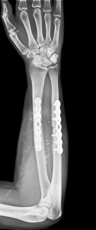
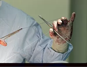
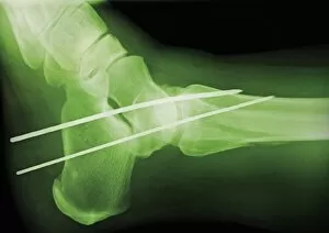
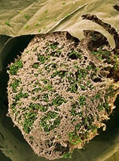
"Fixation: Restoring Strength and Stability through Precision" In the realm of medicine, fixation plays a vital role in mending broken bones and rectifying skeletal anomalies. Picture No. 10899510 captures the essence of this intricate process, showcasing the remarkable journey towards healing. Pinned broken arm, X-ray C017 / 7869 reveals the meticulous work done by skilled surgeons to align fractured bones with precision. Alongside it, Jewish factories on Palestine's Plain Sharon coast symbolize resilience and determination in overcoming adversity. The artwork C016 / 6994 showcases Kirschner wires - delicate tools used to stabilize fractures - resembling an artist's masterpiece. These wires serve as silent heroes behind successful recoveries. Scoliosis treatment is depicted in X-ray C017 / 7156, highlighting how fixation aids in correcting spinal deformities and restoring balance to one's posture. This procedure brings hope for those affected by this condition. Pinned jaw fracture, X-ray C017 / 7982 demonstrates how fixation techniques enable patients to regain their ability to speak and chew without discomfort or limitations. It exemplifies the power of medical advancements that restore normalcy in our lives. X-ray images like C017/7978 reveal pinned broken ankles undergoing fixation procedures – a testament to modern orthopedic practices that allow individuals to walk again confidently after enduring such injuries. Knuckle replacement showcased in X-rays C017/7971 & C017/7970 signifies innovation at its finest; these procedures offer renewed dexterity for those suffering from debilitating joint conditions while preserving mobility for everyday tasks. Furthermore, Pinned fractured elbow displayed in X-ray C017/7969 portrays how precise fixation methods can mend complex fractures effectively—restoring functionality while minimizing long-term complications. Beyond human anatomy lies another form of "fixation": nitrogen-fixing molybdenum iron enzyme (Nif) found within nature itself.