Fraud Blocker Skip to main content

Fifties Collection (page 100)

Step back in time to the fifties, a decade filled with iconic moments and timeless charm

Background imageFifties Collection: Broken elbow, X-ray C017 / 7583

Broken elbow, X-ray C017 / 7583
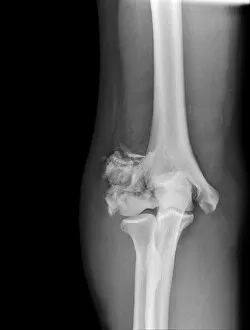
Broken elbow. X-ray of the arm of a 55 year old male patient with fractures to the elbow and radius (upper lower arm bone)

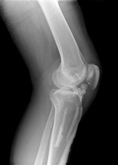
Background imageFifties Collection: Melorheostosis of the knee, X-ray C017 / 7146

Melorheostosis of the knee, X-ray C017 / 7146
Melorheostosis of the knee. X-ray of left knee of a 52 year old male patient showing damage (white area at centre) caused by melorheostosis

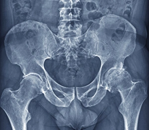
Background imageFifties Collection: Osteoarthritis of the hip, X-ray F006 / 3744

Osteoarthritis of the hip, X-ray F006 / 3744
Osteoarthritis of the hip. Coloured frontal X-ray (front view) of the pelvis in a 59 year old patient showing osteoarthritis of both hip joints

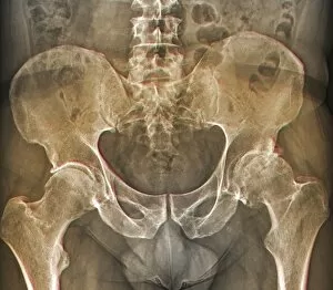
Background imageFifties Collection: Osteoarthritis of the hip, X-ray F006 / 3745

Osteoarthritis of the hip, X-ray F006 / 3745
Osteoarthritis of the hip. Coloured frontal X-ray (front view) of the pelvis in a 59 year old patient showing osteoarthritis of both hip joints

Background imageFifties Collection: Bladder stones, ultrasound scan C017 / 7262

Bladder stones, ultrasound scan C017 / 7262
Bladder stones. Ultrasound scan of the bladder of a 58 year old male showing bladder stones (bright white). Bladder stones (cystoliths)

Background imageFifties Collection: Melorheostosis of the knee, X-ray C017 / 7145

Melorheostosis of the knee, X-ray C017 / 7145
Melorheostosis of the knee. X-ray of left knee of a 52 year old male patient showing damage (white area to bottom left of knee cap) caused by melorheostosis

Background imageFifties Collection: Bladder stones, ultrasound scan C017 / 7263

Bladder stones, ultrasound scan C017 / 7263
Bladder stones. Ultrasound scan of the bladder of a 58 year old male showing bladder stones (bright white). Bladder stones (cystoliths)

Background imageFifties Collection: Melorheostosis of the knee, X-ray C017 / 7144

Melorheostosis of the knee, X-ray C017 / 7144
Melorheostosis of the knee. X-ray of left knee of a 52 year old male patient showing damage (white area to bottom left of knee cap) caused by melorheostosis

Background imageFifties Collection: Archaeon ribosome, molecular model F006 / 9328

Archaeon ribosome, molecular model F006 / 9328
Archaeon ribosome. Molecular model showing the structure of a 50S (large) ribosome from the archaeon Haloarcula marismortui. Ribosomes are composed of protein and RNA (ribonucleic acid)

Background imageFifties Collection: Dislocated finger, X-ray C017 / 7183

Dislocated finger, X-ray C017 / 7183
Dislocated finger. X-ray of the left hand of a 51 year old female patient with a dislocated middle finger. Once the bones have been manipulated back into place treatment is with immobilisation

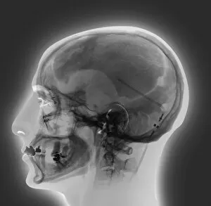
Background imageFifties Collection: Ventricular shunt, X-ray

Ventricular shunt, X-ray
Ventricular shunt. X-ray of the head of a 52-year-old patient with hydrocephalus, showing the position of a ventricular shunt (dark lines, right) and its external valve (round, centre right)

Background imageFifties Collection: Atherosclerosis, 3D CT scan

Atherosclerosis, 3D CT scan
Atherosclerosis. 3D computed tomography (CT) angiogram of the main blood vessels of the lower body a 58-year-old man, showing numerous atheromatous plaques (light, centre) on the vessel walls

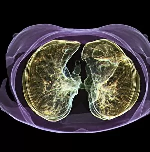
Background imageFifties Collection: Healthy lungs, 3D CT scan C016 / 6504

Healthy lungs, 3D CT scan C016 / 6504
Healthy lungs. Coloured 3D computed tomography (CT) scan of the chest of a 54 year old patient showing healthy lungs. At centre is the aortic arch (red), where the aorta, the bodys main artery

Background imageFifties Collection: Replacement ankle joint, X-ray C016 / 6583

Replacement ankle joint, X-ray C016 / 6583
Replacement ankle joint. Coloured X-ray of the ankles of a 59 year old patient showing a prosthetic ankle joint between the left tibia (shin bone) and talus (ankle bone)

Background imageFifties Collection: Pinned thigh fracture, X-ray C016 / 6563

Pinned thigh fracture, X-ray C016 / 6563
Pinned thigh fracture. Coloured X-ray of the fractured femur (thigh bone) of a 52 year old patient. The broken bone has been fixed internally with a metal rod

Background imageFifties Collection: Healthy lungs, 3D CT scan C016 / 6503

Healthy lungs, 3D CT scan C016 / 6503
Healthy lungs. Coloured 3D computed tomography (CT) scan of the chest of a 54 year old patient showing healthy lungs. At centre is the aortic arch (red), where the aorta, the bodys main artery

Background imageFifties Collection: Healthy lungs, 3D CT scan C016 / 6502

Healthy lungs, 3D CT scan C016 / 6502
Healthy lungs. Coloured 3D computed tomography (CT) scan of the chest of a 54 year old patient showing healthy lungs

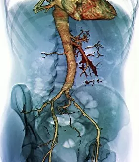
Background imageFifties Collection: Abdominal aorta, 3D CT scan C016 / 6542

Abdominal aorta, 3D CT scan C016 / 6542
Abdominal aorta. Coloured 3D computed tomography (CT) scan of the abdominal aorta of a 56 year old patient. The aorta is the bodys main artery. It carries blood from the heart (at top) around the body

Background imageFifties Collection: Pinned thigh fracture, X-ray C016 / 6564

Pinned thigh fracture, X-ray C016 / 6564
Pinned thigh fracture. Coloured X-ray of the fractured femur (thigh bone) of a 52 year old patient. The broken bone has been fixed internally with a metal rod

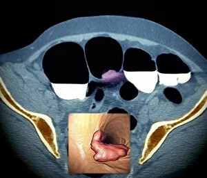
Background imageFifties Collection: Colon cancer, barium contrast CT scan

Colon cancer, barium contrast CT scan
Colon cancer. Coloured barium contrast computed tomography (CT) scan, and 3D CT colonoscopy image (inset), of an axial section through the sigmoid colon (large intestine) of a 54-year-old patient

Background imageFifties Collection: Colon cancer, 3D colonoscopy image

Colon cancer, 3D colonoscopy image
Colon cancer. 3D colonoscopy image showing the location of a cancerous growth (centre right) in the colon of a 59-year-old patient. Here, the growth has caused a narrowing (stenosis) of the colon

Background imageFifties Collection: Narrowed aorta, 3D CT scans

Narrowed aorta, 3D CT scans
Narrowed aorta. Coloured 3D computed tomography (CT) scans of the heart (centre of each scan) and major blood vessels of a 50-year-old patient

Background imageFifties Collection: Colon polyp, 3D colonoscopy image

Colon polyp, 3D colonoscopy image
Colon polyp. Coloured 3D computed tomography (CT) colonoscopy image of a pedunculated (stalked) polyp in the sigmoid colon (large intestine) of a 50-year-old patient

Background imageFifties Collection: Stroke, MRI scan

Stroke, MRI scan
Stroke. Coloured magnetic resonance imaging (MRI, left) and computed tomography (CT, right) scans of axial sections through the brain of a 58-year-old patient

Background imageFifties Collection: Berry aneurysm, MRI scan

Berry aneurysm, MRI scan
Stroke. Coloured computed tomography (CT) scan of an axial section through the brain of a 58-year-old patient, showing the damage (dark) caused 24 hours after a cerebral infarction

Background imageFifties Collection: Stroke, CT scan

Stroke, CT scan
Stroke. Coloured computed tomography (CT) scan of an axial section through the brain of a 58-year-old patient, showing the damage (purple) caused 24 hours after a cerebral infarction

Background imageFifties Collection: Colon cancer, CT scans

Colon cancer, CT scans
Colon cancer. 2D computed tomography (CT) rendering (left) and 3D colonoscopy image (right) showing the location of a cancerous growth in the colon of a 59-year-old patient

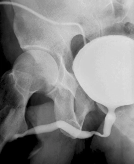
Background imageFifties Collection: Voiding cystourethrogram, X-rays C018 / 0577

Voiding cystourethrogram, X-rays C018 / 0577
Voiding cystourethrogram. X-ray of a 54 year old male patient undergoing a voiding cystourethrogram. This procedure involves liquid containing a radio-opaque dye being placed in the bladder via a

Background imageFifties Collection: Voiding cystourethrogram, X-rays C018 / 0578

Voiding cystourethrogram, X-rays C018 / 0578
Voiding cystourethrogram. Coloured X-ray of a 54 year old male patient undergoing a voiding cystourethrogram. This procedure involves liquid containing a radio-opaque dye being placed in the bladder

Background imageFifties Collection: Liver cancer, CT scan C018 / 0461

Liver cancer, CT scan C018 / 0461
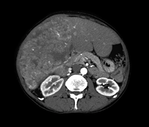
Liver cancer. Coloured computed tomography (CT) scan of the abdomen of a 50 year old alcoholic patient with liver cancer. The liver (orange) is enlarged due to the presence of the tumour

Background imageFifties Collection: Liver cancer, CT scan C018 / 0462

Liver cancer, CT scan C018 / 0462
Liver cancer. Computed tomography (CT) scan of the abdomen of a 50 year old alcoholic patient with liver cancer. The liver (left to upper centre) is enlarged due to the presence of the tumour

Background imageFifties Collection: Liver cancer, CT scan C018 / 0460

Liver cancer, CT scan C018 / 0460
Liver cancer. Coloured computed tomography (CT) scan of the abdomen of a 50 year old alcoholic patient with liver cancer. The liver (orange) is enlarged due to the presence of the tumour

Background imageFifties Collection: Kidney infection, CT scan C018 / 0473

Kidney infection, CT scan C018 / 0473
Kidney infection. Coloured computed tomography (CT) scans of the abdomen of a 57 year old patient with a kidney infection

Background imageFifties Collection: Early-onset Alzheimers disease, MRI scan

Early-onset Alzheimers disease, MRI scan
Early-onset Alzheimers disease. Magnetic resonance imaging (MRI) scans of the brain of a 53 year old patient with early-onset Alzheimers disease

Background imageFifties Collection: Voiding cystourethrogram, X-rays C018 / 0579

Voiding cystourethrogram, X-rays C018 / 0579
Voiding cystourethrogram. Coloured X-ray of a 54 year old male patient undergoing a voiding cystourethrogram. This procedure involves liquid containing a radio-opaque dye being placed in the bladder

Background imageFifties Collection: Broken elbow, X-ray C017 / 7584

Broken elbow, X-ray C017 / 7584
Broken elbow. X-ray of the arm of a 55 year old male patient with fractures to the elbow and radius (right-most lower arm bone)

Background imageFifties Collection: Bladder stones, ultrasound scan C017 / 7261

Bladder stones, ultrasound scan C017 / 7261
Bladder stones. Ultrasound scan of the bladder of a 58 year old male showing bladder stones (bright white). Bladder stones (cystoliths)

Background imageFifties Collection: Arthritic hands, X-ray C017 / 7180

Arthritic hands, X-ray C017 / 7180
Arthritic hands, X-ray

Background imageFifties Collection: Fractured vertebra, X-ray C017 / 7585

Fractured vertebra, X-ray C017 / 7585
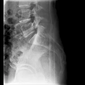
Fractured vertebra. X-ray of the lower spine of a 50 year old male with a fracture to the L1 lumbar vertebra (top centre)

Background imageFifties Collection: Nepalese man C016 / 5891

Nepalese man C016 / 5891
Nepalese man. A Bontava Kiranti of Nepal, aged 55 (circa 1840). Lithograph, Plate 32, from the scrapbook collection made by Hodgson

Background imageFifties Collection: Artificial urinary sphincter, X-ray C016 / 6609

Artificial urinary sphincter, X-ray C016 / 6609
Artificial urinary sphincter. Coloured X-ray of the pelvis of a 55 year old female patient showing a prosthetic urinary sphincter (AUS, beige at centre)

Background imageFifties Collection: Artificial urinary sphincter, X-ray C016 / 6610

Artificial urinary sphincter, X-ray C016 / 6610
Artificial urinary sphincter. Coloured X-ray of the pelvis of a 55 year old female patient showing a prosthetic urinary sphincter (AUS, beige at centre)

Background imageFifties Collection: Artificial urinary sphincter, X-ray C016 / 6608

Artificial urinary sphincter, X-ray C016 / 6608
Artificial urinary sphincter. Coloured X-ray of the pelvis of a 55 year old female patient showing a prosthetic urinary sphincter (AUS, beige at centre)

Background imageFifties Collection: Knee realignment surgery, X-ray C016 / 6604

Knee realignment surgery, X-ray C016 / 6604
Knee alignment surgery. Coloured X-ray of the knee of a 55 year old overweightpatient that has undergone a wedge osteotomy to correct the alignment of the knee joint

Background imageFifties Collection: Knee realignment surgery, X-ray C016 / 6601

Knee realignment surgery, X-ray C016 / 6601
Knee alignment surgery. Coloured X-ray of the knee of a 55 year old overweightpatient that has undergone a wedge osteotomy to correct the alignment of the knee joint

Background imageFifties Collection: Knee realignment surgery, X-ray C016 / 6603

Knee realignment surgery, X-ray C016 / 6603
Knee alignment surgery. Coloured X-ray of the knee of a 55 year old overweightpatient that has undergone a wedge osteotomy to correct the alignment of the knee joint

Background imageFifties Collection: Knee realignment surgery, X-ray C016 / 6602

Knee realignment surgery, X-ray C016 / 6602
Knee alignment surgery. X-ray of the knee of a 55 year old overweightpatient that has undergone a wedge osteotomy to correct the alignment of the knee joint

Background imageFifties Collection: Knee realignment surgery, X-ray C016 / 6599

Knee realignment surgery, X-ray C016 / 6599
Knee alignment surgery. Coloured X-ray of the knee of a 55 year old overweightpatient that has undergone a wedge osteotomy to correct the alignment of the knee joint


All Professionally Made to Order for Quick Shipping

Step back in time to the fifties, a decade filled with iconic moments and timeless charm. In 1953, Her Majesty Queen Elizabeth II ascended to the throne, marking the beginning of an era that would shape history. Meanwhile, at The Locarno Dance Hall in Sauchiehall Street, Glasgow, young couples swayed to the rhythm of love under shimmering lights. The passing out parade at RAF Cranwell showcased the bravery and dedication of our servicemen as they embarked on their noble journey. And who could forget the Ford Thunderbird 1955 Blue & white cruising down sun-kissed streets, capturing hearts with its sleek design? Music echoed through every corner as Billy Fury took center stage in 1958, captivating audiences with his electrifying performances. On two wheels roared the Triumph 650cc Thunderbird 1952 Blue motorcycle - a symbol of freedom and adventure. Sports enthusiasts rejoiced as Partick Thistle FC proudly lifted the Scottish FA Cup in 1954 - a moment etched forever in football history. Willys Jeep and Land Rover Series 1 conquered rugged terrains effortlessly while Vincent Rapide C 1000cc dominated roads with its power. Even racing legend Stirling Moss found solace over a cup of tea amidst his adrenaline-fueled pursuits. And who could resist falling head over heels for the charming Austin-Healey Frogeye Sprite Mk1? They were an enchanting time when dreams soared high and possibilities seemed endless. It was an era defined by elegance, resilience, and innovation - where traditions met modernity seamlessly. So let us embrace nostalgia's warm embrace as we reminisce about this remarkable decade that shaped culture and left an indelible mark on our collective memory.