Fraud Blocker Skip to main content

Fibres Collection (page 7)

"Fibres: Unraveling the Intricacies of the Human Brain and Beyond" Exploring the intricate network of brain fibres through DTI MRI scans (C017 / 7099

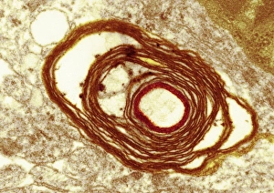
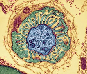
Background imageFibres Collection: Myelin surrounding a nerve axon, TEM

Myelin surrounding a nerve axon, TEM
Myelin surrounding a nerve axon, coloured transmission electron micrograph (TEM). The concentric round rings are the sheets of a Schwann cells myelin membrane (brown rings)

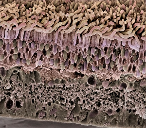
Background imageFibres Collection: Retina, SEM

Retina, SEM
Retina. Coloured scanning electron micrograph (SEM) of a section through a freeze-fractured retina, showing the light-sensitive rods and cones and their associated neurones

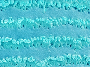
Background imageFibres Collection: Lens fibre of an eye

Lens fibre of an eye
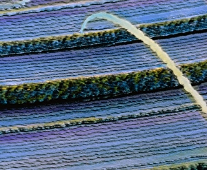
Lens fibre from an eye. Coloured scanning electron micrograph (SEM) of the fibre-like cells that form the crystalline lens of the eye

Background imageFibres Collection: Cornea, SEM

Cornea, SEM
Cornea. Coloured scanning electron micrograph (SEM) of a section through a cornea, the transparent region on the outer surface of the eye. The front of the eye is at top

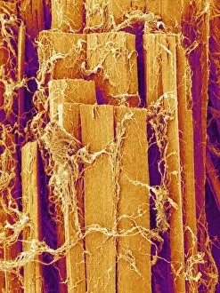
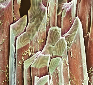
Background imageFibres Collection: Skeletal muscle fibre

Skeletal muscle fibre. Coloured scanning electron micrograph (SEM) of skeletal muscle fibre. This type of muscle is striated

Background imageFibres Collection: Smooth muscle, SEM

Smooth muscle, SEM
Smooth muscle. Coloured scanning electron micrograph of smooth muscle from the trachea (windpipe). Smooth muscle is not under voluntary control, unlike skeletal (striated) muscle

Background imageFibres Collection: Contracted artery, TEM

Contracted artery, TEM
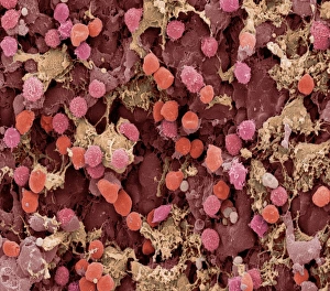
Contracted elastic artery. Coloured transmission electron micrograph (TEM) of a section through an artery. Red blood cells (erythrocytes) are seen in the lumen (top centre)

Background imageFibres Collection: Somatic peripheral nervous system

Somatic peripheral nervous system
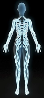
Peripheral nervous system. Computer artwork of the main nerves of the somatic part of the peripheral nervous system. The peripheral nervous system does not include the brain and spinal cord

Background imageFibres Collection: Surgical face mask, SEM

Surgical face mask, SEM
Surgical face mask, coloured scanning electron micrograph (SEM). This mask is made of the plant material cellulose, which is biodegradable

Background imageFibres Collection: Straw particleboard

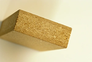
Straw particleboard. Piece of particleboard made from wheat straw. Particles of straw are produced by milling bales of waste straw, and then bound by a resin to produce a wood-like building material

Background imageFibres Collection: Banknote security

Banknote security. Euro banknote in ultraviolet light. This highlights security features intended to prevent fraud. Fluorescent ink and strips glow bright blue and red under ultraviolet light

Background imageFibres Collection: False-colour SEM of cells from lens of hu

False-colour SEM of cells from lens of hu

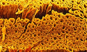
Background imageFibres Collection: Myelinated nerve fibres, SEM

Myelinated nerve fibres, SEM
Myelinated nerve fibres. Coloured scanning electron micrograph (SEM) of a section through a bundle of nerve fibres containing a blood vessel (orange)

Background imageFibres Collection: Neurovascular bundle, TEM

Neurovascular bundle, TEM
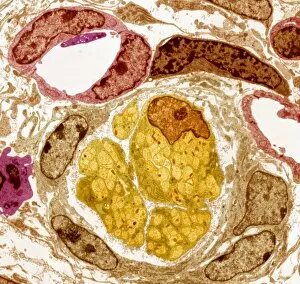
Neurovascular bundle. Coloured transmission electron micrograph (TEM) of a cross section through a neurovascular bundle (NVB)

Background imageFibres Collection: Nerve fibre node, TEM

Nerve fibre node, TEM
Nerve fibre node. Coloured transmission electron micrograph (TEM) of a cross-section through a nerve fibre (axon) at a node of Ranvier

Background imageFibres Collection: Nerve fibres, TEM

Nerve fibres, TEM
Nerve fibres. Coloured transmission electron micrograph (TEM) of a section through non- myelinated nerve fibres (blue). The fibres are arranged into bundles surrounded by the cytoplasm of Schwann

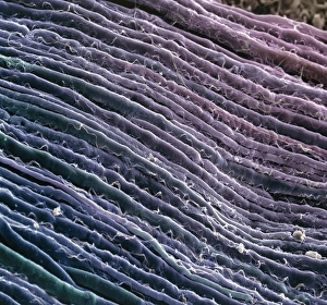
Background imageFibres Collection: Nerve fibres

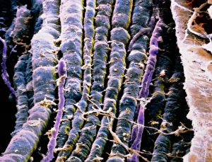
Nerve fibres. Coloured scanning electron micrograph (SEM) of parallel myelinated nerve fibres in the spinal cord. Each fibre consists of a nerve cell axon, the output process of a nerve cell

Background imageFibres Collection: Nerve fibre, SEM

Nerve fibre, SEM
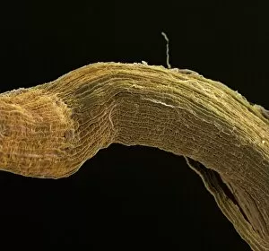
Nerve fibres. Coloured scanning electron micrograph (SEM) of nerve fibres (brown). A group of nerves such as this is known as a fasciculus

Background imageFibres Collection: Coloured SEM of some nerve fibres

Coloured SEM of some nerve fibres
Nerve fibres. Coloured scanning electron micrograph (SEM) of some nerve fibres. A group of nerves such as this is known as a fasciculus

Background imageFibres Collection: False-colour SEM of a bundle of motor nerve fibres

False-colour SEM of a bundle of motor nerve fibres
Nerve fibres. False-colour scanning electron micrograph of a bundle of motor nerve fibres (blue). The bundle as a whole is surrounded by a cylindrical sheath of connective tissue

Background imageFibres Collection: Ilustration of an oligodendrocyte

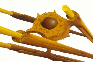
Ilustration of an oligodendrocyte

Background imageFibres Collection: Lymph node, SEM

Lymph node, SEM
Lymph node. Coloured scanning electron micrograph (SEM) of a freeze fracture section through a lymph node, showing part of the medullary sinus

Background imageFibres Collection: Blood vessels, light micrograph

Blood vessels, light micrograph
Blood vessels. Light micrograph of a section through tissue showing an artery (bottom left) and a vein (top right). Two nerve fibres (dark blue) are also seen

Background imageFibres Collection: Muscular artery

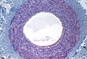
Muscular artery. Light micrograph of a transverse section through a muscular artery. This blood vessel has a thick, muscular wall that enables it to carry blood under pressure

Background imageFibres Collection: Muscle fibres, SEM

Muscle fibres, SEM
Skeletal muscle fibres. Coloured scanning electron micrograph (SEM) of a freeze-fractured bundle of skeletal (or striated) muscle fibres

Background imageFibres Collection: Tendon fibres, TEM

Tendon fibres, TEM
Tendon fibres, coloured transmission electron micrograph (TEM). A tendon is made up of parallel bundles of collagen fibres

Background imageFibres Collection: Skeletal muscle, SEM

Skeletal muscle, SEM
Skeletal muscle. Scanning electron micrograph (SEM) of a transverse section through skeletal muscle. The many muscle fibres (red) are seen, along with the elastic sheath (epimysium)


All Professionally Made to Order for Quick Shipping

"Fibres: Unraveling the Intricacies of the Human Brain and Beyond" Exploring the intricate network of brain fibres through DTI MRI scans (C017 / 7099). The mesmerizing artwork depicting white matter fibres and their connection to the brain (C015 / 1930). Delving deeper into understanding brain fibres with DTI MRI scans (C017 / 7035). Illuminating neurons and astrocytes through immunofluorescent LM, revealing the complexity of brain fibres. Peering into the white matter fibres of the human brain, unlocking its mysteries (C014 / 5666). Witnessing nerve cells regenerate under a TEM microscope, showcasing nature's resilience. Marveling at the intricate structure of a lime tree stem captured in a light micrograph. Discovering beauty in unexpected places - castor oil stem captured in a mesmerizing light micrograph. Unveiling myelination's role in enhancing nerve fibre function through TEM imaging. Witnessing cell division unfold under fluorescent micrographs, highlighting life's continuous renewal process. Celebrating nature's ability to heal as we observe regenerating nerve cells under TEM microscopy once again. Exploring further into white matter fibres within our complex human brains (C014/5668). In this captivating journey, we unravel the wonders – from delicate neural connections within our brains to resilient plant stems that provide sustenance for life itself.