
Fallopian Tube Collection
"The Incredible Journey: Exploring the Marvels of the Fallopian Tube" Embark on a captivating visual voyage through the intricate world of human reproduction


All Professionally Made to Order for Quick Shipping
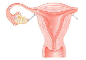
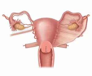
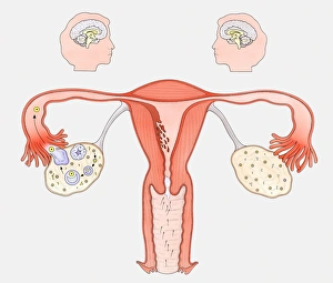
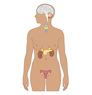
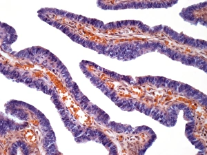
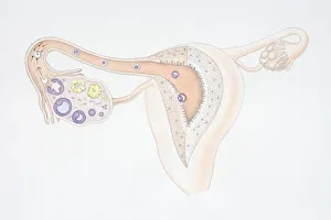
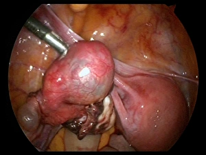
"The Incredible Journey: Exploring the Marvels of the Fallopian Tube" Embark on a captivating visual voyage through the intricate world of human reproduction. A digital illustration unveils the awe-inspiring journey of a fertilized egg within the fallopian tube, where life begins its miraculous formation. Delve deeper into this fascinating realm with a cross-sectional biomedical illustration showcasing the female reproductive system. Witness an anterior view of a normal uterus, ovaries, fallopian tubes, and broad ligament – nature's masterpiece crafted for nurturing new life. In contrast, another cross-sectional view reveals an intimate perspective of a uterus afflicted by fibroids. This depiction sheds light on various conditions that can affect women's health and fertility. Unraveling further mysteries lies a diagram illustrating how female sexual organs interact with our brain. On one side, witness the intricate dance of hormones orchestrating the normal reproductive cycle; while on the other side, discover how contraceptive pills influence this delicate equilibrium. A black and white illustration showcases naked womanhood in all its glory – unveiling not only her uterus but also her fallopian tubes, ovary, and vagina. It celebrates both beauty and functionality in perfect harmony. Witness medical marvels as we explore endoscope views revealing uterine wonders from within. Uterus endoscope view C017/6805 offers an up-close encounter with this remarkable organ that holds immense significance in creating life. Discover yet another innovation designed to empower women's choices – delve into a biomedical illustration depicting vaginal rings positioned strategically within their intended environment. This revolutionary method revolutionizes contraception options for modern-day females. The complexity doesn't stop there. Cross-section illustrations unveil secrets held by adult females' endocrine systems - intricately woven networks responsible for regulating hormonal balance throughout their lives. Microscopic wonders come alive through light micrograph F006/9799 capturing mesmerizing details of fallopian tubes at cellular levels - a testament to the intricate design that facilitates conception.