Fraud Blocker Skip to main content

Erythrocyte Collection (page 5)

"Erythrocyte: The Lifeline of Blood" Erythrocytes, commonly known as red blood cells, play a crucial role in our circulatory system

Background imageErythrocyte Collection: Red blood cells, artwork C013 / 5932

Red blood cells, artwork C013 / 5932
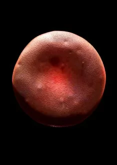
Red blood cells (erythrocytes), computer artwork

Background imageErythrocyte Collection: Malarial blood cell, SEM C017 / 8307

Malarial blood cell, SEM C017 / 8307
Malarial blood cell. Coloured scanning electron micrograph (SEM) of an erythrocyte (red blood cell) infected with Plasmodium parasites, the cause of malaria

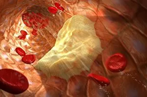
Background imageErythrocyte Collection: Atheromatous artery, artwork C015 / 6445

Atheromatous artery, artwork C015 / 6445
Atheromatous artery. Computer artwork showing the interior of an artery affected by atherosclerosis. Atherosclerosis is narrowing of an artery (stenosis) due to fatty deposits (beige)

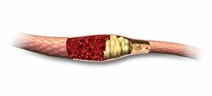
Background imageErythrocyte Collection: Atheromatous artery, artwork C015 / 6446

Atheromatous artery, artwork C015 / 6446
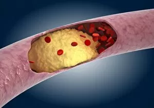
Atheromatous artery. Cutaway computer artwork showing the interior of an artery affected by atherosclerosis. Atherosclerosis is narrowing of an artery (stenosis) due to fatty deposits (beige)

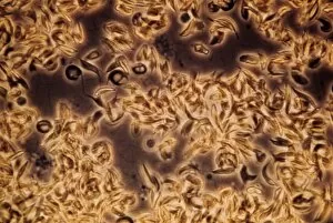
Background imageErythrocyte Collection: Sickle cell anaemia, light micrograph C015 / 6395

Sickle cell anaemia, light micrograph C015 / 6395
Sickle cell anaemia. Light micrograph of a blood smear showing normally rounded red blood cells that have become curved, elongated sickle-shaped cells

Background imageErythrocyte Collection: Kidney glomerulus, TEM C016 / 5831

Kidney glomerulus, TEM C016 / 5831
Kidney glomerulus. Transmission electron micrograph (TEM) of a section through a renal glomerulus. At centre left is a capillary with a red blood cell (black) in its lumen

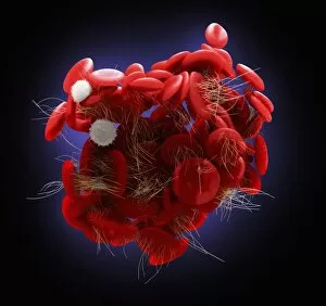
Background imageErythrocyte Collection: Blood clot, artwork C016 / 4618

Blood clot, artwork C016 / 4618
Blood clot, computer artwork. Red blood cells (erythrocytes) are trapped within a fibrin protein mesh (cream). The fibrin mesh is formed in response to chemicals secreted by platelets (white)

Background imageErythrocyte Collection: Red blood cells, artwork C016 / 4627

Red blood cells, artwork C016 / 4627
Red blood cells, computer artwork. Red blood cells are biconcave, giving them a large surface area for gas exchange, and highly elastic, enabling them to pass through narrow capillary vessels

Background imageErythrocyte Collection: Red blood cells, artwork C016 / 4626

Red blood cells, artwork C016 / 4626
Red blood cells, computer artwork. Red blood cells are biconcave, giving them a large surface area for gas exchange, and highly elastic, enabling them to pass through narrow capillary vessels

Background imageErythrocyte Collection: Red blood cell, artwork C016 / 4622

Red blood cell, artwork C016 / 4622
Red blood cell, computer artwork. Red blood cells are biconcave, giving them a large surface area for gas exchange, and highly elastic, enabling them to pass through narrow capillary vessels

Background imageErythrocyte Collection: Red blood cell, artwork C016 / 4623

Red blood cell, artwork C016 / 4623
Red blood cell, computer artwork. Red blood cells are biconcave, giving them a large surface area for gas exchange, and highly elastic, enabling them to pass through narrow capillary vessels

Background imageErythrocyte Collection: Red blood cell, artwork C016 / 4624

Red blood cell, artwork C016 / 4624
Red blood cell, computer artwork. Red blood cells are biconcave, giving them a large surface area for gas exchange, and highly elastic, enabling them to pass through narrow capillary vessels

Background imageErythrocyte Collection: Blood clot, artwork C016 / 4620

Blood clot, artwork C016 / 4620
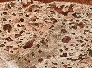
Fortification agate. Section through a fortification agate from Scurdie Ness, Scotland, UK. Agate is an aggregate of different forms of silica (SiO2) within a geode

Background imageErythrocyte Collection: Erythropoietin hormone complex C016 / 4461

Erythropoietin hormone complex C016 / 4461
Erythropoietin hormone complex. Computer model showing the secondary structure of a molecule of the human hormone Erythropoietin (EPO), complexed with an erythropoetin receptor molecule

Background imageErythrocyte Collection: Erythropoietin hormone complex C016 / 4458

Erythropoietin hormone complex C016 / 4458
Erythropoietin hormone complex. Computer model showing the secondary structure of a molecule of the human hormone Erythropoietin (EPO), complexed with an erythropoetin receptor molecule

Background imageErythrocyte Collection: Erythropoietin hormone complex C016 / 4451

Erythropoietin hormone complex C016 / 4451
Erythropoietin hormone complex. Computer model showing the secondary structure of a molecule of the human hormone Erythropoietin (EPO), complexed with an erythropoetin receptor molecule

Background imageErythrocyte Collection: Blood cells, SEM C016 / 3101

Blood cells, SEM C016 / 3101
Blood cells. Coloured scanning electron micrograph (SEM) of a variety of different white blood cells (leucocytes, blue/purple) with two red blood cells (erythrocytes)

Background imageErythrocyte Collection: Blood cells, SEM C016 / 3100

Blood cells, SEM C016 / 3100
Blood cells. Coloured scanning electron micrograph (SEM) of a variety of different white blood cells (leucocytes, yellow/green) with two red blood cells (erythrocytes)

Background imageErythrocyte Collection: Blood cells, SEM C016 / 3096

Blood cells, SEM C016 / 3096
Blood cells. Coloured scanning electron micrograph of a lymphocyte white blood cell (left) and a red blood cell (erythrocyte, right)

Background imageErythrocyte Collection: Red blood cells, light micrograph C016 / 3036

Red blood cells, light micrograph C016 / 3036
Red blood cells. Differential interference contrast (DIC) micrograph of red blood cells (erythrocytes). Red blood cells are biconcave, giving them a large surface area for gas exchange

Background imageErythrocyte Collection: Malaria parasite in red blood cell C016 / 2444

Malaria parasite in red blood cell C016 / 2444
Malaria parasite in red blood cell. Light micrograph of a red blood cell (erythrocyte) infected with a Plasmodim vivax protozoan. This parasite causes recurring malaria in humans

Background imageErythrocyte Collection: Malaria parasite in red blood cell C016 / 2443

Malaria parasite in red blood cell C016 / 2443
Malaria parasite in red blood cell. Light micrograph of a red blood cell (erythrocyte) infected with a Plasmodim vivax protozoan. This parasite causes recurring malaria in humans

Background imageErythrocyte Collection: Malaria parasite in red blood cell C016 / 2442

Malaria parasite in red blood cell C016 / 2442
Malaria parasite in red blood cell. Light micrograph of a red blood cell (erythrocyte) infected with a Plasmodim vivax protozoan. This parasite causes recurring malaria in humans

Background imageErythrocyte Collection: Erythropoietin molecule C013 / 8891

Erythropoietin molecule C013 / 8891
Erythropoietin molecule (EPO), molecular model. EPO is a glycoprotein that is produced by the kidneys in response to low blood oxygen levels

Background imageErythrocyte Collection: Erythropoietin hormone complex

Erythropoietin hormone complex. Computer model showing the secondary structure of a molecule of the human hormone Erythropoietin (EPO), complexed with an erythropoetin receptor molecule

Background imageErythrocyte Collection: Red blood cells (erythrocytes), artwork C013 / 8812

Red blood cells (erythrocytes), artwork C013 / 8812
Red blood cells (erythrocytes), computer artwork

Background imageErythrocyte Collection: Anaemia, conceptual image C013 / 7788

Anaemia, conceptual image C013 / 7788
Anaemia, conceptual image. Computer artwork of anaemic red blood cells, which are no longer red as they have lost their haemoglobin and therefore their ability to carry iron

Background imageErythrocyte Collection: Blood clot C013 / 7129

Blood clot C013 / 7129
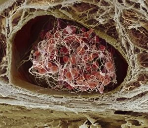
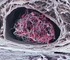
Blood clot. Coloured scanning electron micrograph (SEM) of a blood clot (centre) forming in connective tissue. Red blood cells (erythrocytes)

Background imageErythrocyte Collection: Blood clot C013 / 7128

Blood clot C013 / 7128
Blood clot. Coloured scanning electron micrograph (SEM) of a blood clot (centre) forming in connective tissue. Red blood cells (erythrocytes)

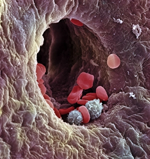
Background imageErythrocyte Collection: Blood-filled artery, SEM C013 / 7117

Blood-filled artery, SEM C013 / 7117
Blood-filled artery. Coloured scanning electron micrograph (SEM) of a section through an artery (dark brown) in muscle tissue (light brown), showing the red blood cells (erythrocytes, centre) inside

Background imageErythrocyte Collection: Blood-filled artery, SEM C013 / 7118

Blood-filled artery, SEM C013 / 7118
Blood-filled artery. Coloured scanning electron micrograph (SEM) of a section through an artery (centre) in muscle tissue, showing the red blood cells (erythrocytes, red) inside

Background imageErythrocyte Collection: Blood-filled artery, SEM C013 / 7116

Blood-filled artery, SEM C013 / 7116
Blood-filled artery. Coloured scanning electron micrograph (SEM) of a section through an artery (dark pink, centre) in muscle tissue (purple), showing the red blood cells (erythrocytes, red) inside

Background imageErythrocyte Collection: Red blood cells, artwork C013 / 4616

Red blood cells, artwork C013 / 4616
Red blood cells. Computer artwork of red blood cells (erythrocytes) flowing through a blood vessel

Background imageErythrocyte Collection: Red blood cells, artwork C013 / 4615

Red blood cells, artwork C013 / 4615
Red blood cells. Computer artwork of red blood cells (erythrocytes) flowing through a blood vessel

Background imageErythrocyte Collection: Atheromatous artery C013 / 4612

Atheromatous artery C013 / 4612
Atheromatous artery. Cutaway computer artwork showing the interior of an artery affected by atherosclerosis, showing red blood cells (red)

Background imageErythrocyte Collection: Blood cells, artwork C013 / 4726

Blood cells, artwork C013 / 4726
Blood cells. Computer artwork of red blood cells (erythrocytes, red), white blood cells (leucocytes, white), and platelets (thrombocytes, yellow)

Background imageErythrocyte Collection: Red blood cells, artwork C013 / 4725

Red blood cells, artwork C013 / 4725
Red blood cells (erythrocytes), computer artwork

Background imageErythrocyte Collection: Anaemia, conceptual image C013 / 4711

Anaemia, conceptual image C013 / 4711
Anaemia, conceptual image. Computer artwork of normal red blood cells (red) and anaemic red blood cells (clear). The anaemic cells are no longer red as they have lost their haemoglobin

Background imageErythrocyte Collection: Blood cells, artwork C013 / 4707

Blood cells, artwork C013 / 4707
Blood cells. Computer artwork of red blood cells (erythrocytes, red), white blood cells (leucocytes, white), and platelets (thrombocytes, yellow)

Background imageErythrocyte Collection: Anaemia, conceptual image C013 / 4709

Anaemia, conceptual image C013 / 4709
Anaemia, conceptual image. Computer artwork of normal red blood cells (red) and anaemic red blood cells (clear). The anaemic cells are no longer red as they have lost their haemoglobin

Background imageErythrocyte Collection: Red blood cells, artwork C013 / 4705

Red blood cells, artwork C013 / 4705
Red blood cells (erythrocytes), computer artwork

Background imageErythrocyte Collection: Red blood cells, artwork C013 / 4706

Red blood cells, artwork C013 / 4706
Red blood cells (erythrocytes), computer artwork

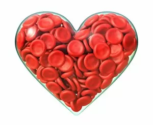
Background imageErythrocyte Collection: Heart care, conceptual image C013 / 4692

Heart care, conceptual image C013 / 4692
Heart care, conceptual image. Computer artwork of red blood cells (erythrocytes) inside heart shapes

Background imageErythrocyte Collection: Heart care, conceptual image C013 / 4691

Heart care, conceptual image C013 / 4691
Heart care, conceptual image. Computer artwork of red blood cells (erythrocytes) inside a heart shape

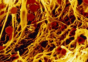
Background imageErythrocyte Collection: False-colour SEM of connective tissue

False-colour SEM of connective tissue
False-colour scanning electron micrograph (SEM) of human connective tissue, showing collagen fibres, which appear as an irregular mass of yellow strands

Background imageErythrocyte Collection: Liver capillary, SEM

Liver capillary, SEM
Liver capillary. Coloured scanning electron micro- graph (SEM) of a freeze-fractured section through liver tissue showing a hepatic capillary, or sinusoid

Background imageErythrocyte Collection: Lung alveoli, SEM

Lung alveoli, SEM
Lung alveoli. Coloured scanning electron micrograph (SEM) of alveoli (air spaces) in the lungs. Blood vessels containing erythrocytes (red blood cells) are also seen

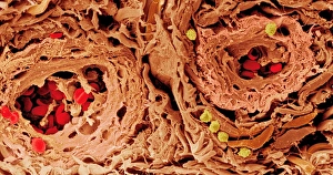
Background imageErythrocyte Collection: Coloured SEM of blood vessels in the skin

Coloured SEM of blood vessels in the skin
Skin blood vessels. Coloured scanning electron micrograph (SEM) of two small blood vessels (far left and upper right) in the dermis of the skin


All Professionally Made to Order for Quick Shipping

"Erythrocyte: The Lifeline of Blood" Erythrocytes, commonly known as red blood cells, play a crucial role in our circulatory system. These tiny, disc-shaped cells are responsible for transporting oxygen from the lungs to every tissue and organ in our body. During menstruation, the uterus lining sheds and erythrocytes come into action. They help deliver necessary nutrients and oxygen to support the regrowth of this lining, ensuring a healthy reproductive cycle. Under scanning electron microscopy (SEM), these remarkable blood cells reveal their intricate structure. Their biconcave shape allows for maximum surface area exposure, facilitating efficient gas exchange within our bodies. The importance of erythrocytes extends beyond just oxygen transport. They also contribute to the complex process of blood coagulation cascade – an essential mechanism that prevents excessive bleeding when injuries occur. Artwork C016/9873 beautifully illustrates this intricate cascade that leads to clot formation. In SEM image C016/9747, we witness a close-up view of a blood clot formed by platelets and fibrin strands working together harmoniously to seal wounds effectively. Human red blood corpuscles captured under SEM showcase their vibrant nature while reminding us of their vital function within our bloodstream - maintaining homeostasis by carrying carbon dioxide back to the lungs for elimination. Even parasites like mouse malaria can be observed through SEM attached to erythrocytes - highlighting how these cells serve as hosts during infection but also become targets for immune responses against such invaders. Blood vessels act as highways where erythrocytes travel tirelessly throughout our bodies. These microscopic highways ensure proper distribution of nutrients and removal of waste products from various tissues they reach. SEM images further unveil intriguing details about erythrocyte behavior; one such example is seen in an image depicting a blood clot forming on plaster's surface – showcasing how these incredible cells respond swiftly when faced with injury or damage outside the body too.