
Endoscopy Collection
"Exploring the Depths: Unveiling the Mysteries of Endoscopy" Endoscopy, a revolutionary medical technique


All Professionally Made to Order for Quick Shipping
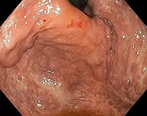
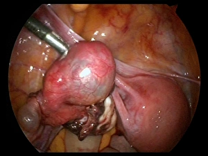
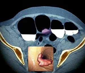
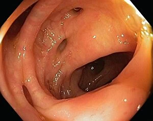
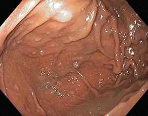
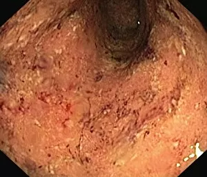
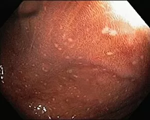
"Exploring the Depths: Unveiling the Mysteries of Endoscopy" Endoscopy, a revolutionary medical technique, allows doctors to delve into the intricate world of our internal organs. With its ability to capture detailed images and provide valuable insights, it has become an indispensable tool in diagnosing and treating various conditions. One area where endoscopy proves invaluable is in examining the intestinal lining. By inserting a flexible tube equipped with a camera, doctors can visualize abnormalities such as ulcerative pancolitis or pouchitis. These conditions affect the colon and can cause discomfort and inflammation. Moving further down the body, endoscope views reveal fascinating glimpses into reproductive health. Ovarian cysts come into focus through C017 / 6800, C017 / 6801, and C017 / 6802 images – providing crucial information for diagnosis and treatment options. Additionally, ectopic pregnancy (C017 / 6804) showcases how this procedure aids in identifying potentially life-threatening situations. Not limited to just female reproductive organs, it also plays a role in exploring other areas like the uterus (C017 / 6805). This view helps detect any abnormalities that may require intervention or monitoring. Venturing beyond reproductive health brings us to gastric antral vascular ectasia (GAVE), which affects blood vessels within the stomach lining (C016 / 8328). Through precise visualization using an endoscope, doctors can accurately diagnose GAVE and devise appropriate treatment plans. The versatility extends even further by encompassing nasal passages and throat examinations. Biomedical illustrations depicting cross-sections highlight how this technique aids in detecting issues within these vital airways. Lastly but significantly important is its role in cancer detection. Colon cancer screenings often involve barium contrast CT scans that utilize endoscopic techniques for accurate imaging results. Endoscopy serves as a window into our bodies' inner workings, allowing medical professionals to diagnose and treat conditions affecting various organs.