Fraud Blocker Skip to main content

Endoscope Collection

"Exploring the Inner World: Unveiling Medical Marvels with Endoscope" Journey into the Unknown

Background imageEndoscope Collection: Ovarian cyst, endoscope view C017 / 6800

Ovarian cyst, endoscope view C017 / 6800
Ovarian cyst. Endoscope (laparascope) view of a cyst (white) on a patients ovary. Ovarian cysts are common in women of child-bearing age

Background imageEndoscope Collection: Uterus, endoscope view C017 / 6805

Uterus, endoscope view C017 / 6805
Uterus. Endoscope (laparascope) view of a healthy uterus (womb, large, upper centre), with the two fallopian tubes (left and right) and an ovary (white, lower right)

Background imageEndoscope Collection: Gastric antral vascular ectasia C016 / 8328

Gastric antral vascular ectasia C016 / 8328
Gastric antral vascular ectasia. Endoscopic view of the antrum (final portion of stomach), affected by gastric antral vascular ectasia (GAVE)

Background imageEndoscope Collection: Colon cancer, endoscopic view C016 / 8308

Colon cancer, endoscopic view C016 / 8308
Colon cancer. Endoscopic view of a malignant (cancerous) adenocarcinoma (upper right) in a patients ascending colon (large intestine)

Background imageEndoscope Collection: Severe gastritis, endoscope view C016 / 3898

Severe gastritis, endoscope view C016 / 3898
Severe gastritis, endoscope view. Gastritis is inflammation of the stomach lining. It causes pain on eating or drinking, and may cause nausea and vomiting

Background imageEndoscope Collection: Cross section biomedical illustration of endoscopy of the nose and throat

Cross section biomedical illustration of endoscopy of the nose and throat

Background imageEndoscope Collection: Cross section biomedical illustration of inside the knee joint during rigid endoscopy procedure

Cross section biomedical illustration of inside the knee joint during rigid endoscopy procedure

Background imageEndoscope Collection: Biomedical illustration of tip of flexible endoscope

Biomedical illustration of tip of flexible endoscope

Background imageEndoscope Collection: Biomedical illustration of rigid endoscope

Biomedical illustration of rigid endoscope

Background imageEndoscope Collection: Detail of human knee showing insertion of arthroscopic instruments

Detail of human knee showing insertion of arthroscopic instruments

Background imageEndoscope Collection: Detail of human knee showing arthroscopic surgical proceedures

Detail of human knee showing arthroscopic surgical proceedures

Background imageEndoscope Collection: Endoscope

Endoscope

Background imageEndoscope Collection: 19th century medical inventions, including early syringes, sphygmomanometer, Letheon ether inhaler

19th century medical inventions, including early syringes, sphygmomanometer, Letheon ether inhaler, ceramic false teeth, endoscope, thermometers, stethoscopes

Background imageEndoscope Collection: Ovarian cyst, endoscope view C017 / 6801

Ovarian cyst, endoscope view C017 / 6801
Ovarian cyst. Endoscope (laparascope) view of a cyst (white) on a patients ovary. Ovarian cysts are common in women of child-bearing age

Background imageEndoscope Collection: Capsule endoscope, artwork F005 / 0186

Capsule endoscope, artwork F005 / 0186
Capsule endoscope, computer artwork

Background imageEndoscope Collection: Ulcerative pancolitis C018 / 0928

Ulcerative pancolitis C018 / 0928
Ulcerative pancolitis. Endoscope view of the appendix aperture in the caecum (start of the large intestine) of an 11 year old patient with ulcerative pancolitis showing inflamed mucosa

Background imageEndoscope Collection: Ovary, endoscope view C017 / 6803

Ovary, endoscope view C017 / 6803
Ovary. Endoscope (laparascope) view of a healthy ovary (white). There are two ovaries in the female body, which release an egg (ovum) during the monthly ovulation cycle

Background imageEndoscope Collection: Ovarian cyst, endoscope view C017 / 6802

Ovarian cyst, endoscope view C017 / 6802
Ovarian cyst. Endoscope (laparascope) view of a cyst (white) on a patients ovary. Ovarian cysts are common in women of child-bearing age

Background imageEndoscope Collection: Ectopic pregnancy, endoscope view C017 / 6804

Ectopic pregnancy, endoscope view C017 / 6804
Ectopic pregnancy. Endoscope (laparascope) view of an ectopic pregnancy in a patients fallopian tube. An ectopic pregnancy (eccysis)

Background imageEndoscope Collection: Pouchitis, endoscope view C015 / 5074

Pouchitis, endoscope view C015 / 5074
Pouchitis, endoscope view. This is the inflammation of an artificial rectum, known as an ileal pouch, formed in patients that have had their large intestine removed

Background imageEndoscope Collection: Thorascopic surgery, artwork C016 / 6842

Thorascopic surgery, artwork C016 / 6842
Thorascopic surgery. Artwork of an overhead view of surgeons carrying out video-assisted thoracoscopic surgery (VATS). This is a form of keyhole (minimally invasive) endoscopic surgery

Background imageEndoscope Collection: Colon cancer, barium contrast CT scan

Colon cancer, barium contrast CT scan
Colon cancer. Coloured barium contrast computed tomography (CT) scan, and 3D CT colonoscopy image (inset), of an axial section through the sigmoid colon (large intestine) of a 54-year-old patient

Background imageEndoscope Collection: Colon cancer, 3D colonoscopy image

Colon cancer, 3D colonoscopy image
Colon cancer. 3D colonoscopy image showing the location of a cancerous growth (centre right) in the colon of a 59-year-old patient. Here, the growth has caused a narrowing (stenosis) of the colon

Background imageEndoscope Collection: Colon polyp, 3D colonoscopy image

Colon polyp, 3D colonoscopy image
Colon polyp. Coloured 3D computed tomography (CT) colonoscopy image of a pedunculated (stalked) polyp in the sigmoid colon (large intestine) of a 50-year-old patient

Background imageEndoscope Collection: Colon cancer, CT scans

Colon cancer, CT scans
Colon cancer. 2D computed tomography (CT) rendering (left) and 3D colonoscopy image (right) showing the location of a cancerous growth in the colon of a 59-year-old patient

Background imageEndoscope Collection: Bowel disease in the colon C016 / 8338

Bowel disease in the colon C016 / 8338
Bowel disease in the colon. Endoscopic view of diverticular disease (diverticulosis) affecting the sigmoid colon. Diverticulosis causes sacs (diverticula)

Background imageEndoscope Collection: Secondary bowel cancer, endoscopic view C016 / 8337

Secondary bowel cancer, endoscopic view C016 / 8337
Secondary bowel cancer. Endoscopic view of the interior of a patients descending duodenum (first part of the small intestine), showing cancer that has spread (metastasised) from the pancreas

Background imageEndoscope Collection: Intestinal lymphoid hyperplasia C016 / 8334

Intestinal lymphoid hyperplasia C016 / 8334
Intestinal lymphoid hyperplasia. Endoscopic view of lymphoid hyperplasia in the terminal ileum - the last part of the small intestine

Background imageEndoscope Collection: Hiatal hernia, endoscopic view C016 / 8330

Hiatal hernia, endoscopic view C016 / 8330
Hiatal hernia. Endoscopic view of a hiatal hernia (gastro-oesophageal prolapse), seen from below, with the endoscope(black) seen in an inverted position inside the stomach

Background imageEndoscope Collection: Hiatal hernia, endoscopic view C016 / 8329

Hiatal hernia, endoscopic view C016 / 8329
Hiatal hernia. Endoscopic view of a hiatal hernia (gastro-oesophageal prolapse), seen from above. A hernia is the protrusion of an organ, or the fascia of an organ

Background imageEndoscope Collection: Hyperplastic polyps in the stomach C016 / 8327

Hyperplastic polyps in the stomach C016 / 8327
Hyperplastic polyps in the stomach. Endoscopic view of small hyperplastic polyps (growths) in the gastric fundus of the stomach

Background imageEndoscope Collection: Major duodenal papilla, endoscopic view C016 / 8324

Major duodenal papilla, endoscopic view C016 / 8324
Major duodenal papilla. Endoscopic view of the interior of a persons small intestine, showing the major duodenal papilla. This structure is where the common bile duct

Background imageEndoscope Collection: Small intestine, endoscopic view C016 / 8323

Small intestine, endoscopic view C016 / 8323
Small intestine. Endoscopic view of the healthy interior of a persons duodenum - the first part of the small intestine, showing the muscular wall

Background imageEndoscope Collection: Lining of the duodenum, endoscopic view C016 / 8321

Lining of the duodenum, endoscopic view C016 / 8321
Lining of the duodenum. Endoscopic view of the lining of the duodenum - the first section of the small intestine - showing lipid (fat, white) filled villi

Background imageEndoscope Collection: Intestinal polyp removal, endoscopic view C016 / 8325

Intestinal polyp removal, endoscopic view C016 / 8325
Intestinal polyp removal. Endoscopic view of a polyp (centre-right) being removed from the sigmoid colon (large intestine). Polyps are benign (non-cancerous) growths that arise from mucous membranes

Background imageEndoscope Collection: Diverticulum of the duodenum C016 / 8320

Diverticulum of the duodenum C016 / 8320
Diverticulum of the duodenum. Endoscope view of a diverticulum in the wall of the descending part of the duodenum - the first section of the small intestine

Background imageEndoscope Collection: Angiodysplasia, endoscopic view C016 / 8318

Angiodysplasia, endoscopic view C016 / 8318
Angiodysplasia. Endoscopic view of the interior of a patients caecum (beginning of the large intestine), showing a small angiodysplasia

Background imageEndoscope Collection: Crohns disease, endoscopic view

Crohns disease, endoscopic view
Crohns disease. Endoscopic view of the interior of a patients neoterminal ileum affected by Crohns disease, showing ulceration (white)

Background imageEndoscope Collection: Ileocaecal valve, endoscopic view C016 / 8317

Ileocaecal valve, endoscopic view C016 / 8317
Ileocaecal valve. Endoscopic view of the healthy interior of a persons ileum (small intestine), showing the ileocaecal valve

Background imageEndoscope Collection: Ulcerative colitis, endoscopic view C016 / 8313

Ulcerative colitis, endoscopic view C016 / 8313
Ulcerative colitis. Endoscopic view of the interior of a patients distal colon (large intestine) affected by ulcerative colitis (UC), showing ulceration (white areas)

Background imageEndoscope Collection: Antrum occult mucosal bleeding C016 / 8314

Antrum occult mucosal bleeding C016 / 8314
Antrum occult mucosal bleeding. Endoscopic view of the interior of a patients antrum (final portion of stomach), showing bleeding (red) of the mucosal lining

Background imageEndoscope Collection: Ulcerative colitis, endoscopic view C016 / 8312

Ulcerative colitis, endoscopic view C016 / 8312
Ulcerative colitis. Endoscopic view of the interior of a patients distal colon (large intestine) affected by ulcerative colitis (UC), showing ulceration (white areas)

Background imageEndoscope Collection: Ulcerative proctitis, endoscopic view C016 / 8311

Ulcerative proctitis, endoscopic view C016 / 8311
Ulcerative colitis. Endoscopic view of the interior of a patients anus, showing ulceration (white areas) cause by ulcerative proctitis

Background imageEndoscope Collection: Ulcerative colitis, endoscopic view C016 / 8310

Ulcerative colitis, endoscopic view C016 / 8310
Ulcerative colitis. Endoscopic view of the interior of a patients colon (large intestine) affected by ulcerative colitis (UC), showing ulceration (white areas)

Background imageEndoscope Collection: Ulcerative colitis, endoscopic view C016 / 8309

Ulcerative colitis, endoscopic view C016 / 8309
Ulcerative colitis. Endoscopic view of the interior of a patients colon (large intestine) affected by ulcerative colitis (UC), showing ulceration (white areas)

Background imageEndoscope Collection: Ulcerative colitis, endoscopic view C016 / 8306

Ulcerative colitis, endoscopic view C016 / 8306
Ulcerative colitis. Endoscopic view of the interior of a patients distal colon (large intestine) affected by ulcerative colitis (UC), showing ulceration (white areas)

Background imageEndoscope Collection: Colon cancer, endoscopic view C016 / 8307

Colon cancer, endoscopic view C016 / 8307
Colon cancer. Endoscopic view of a malignant (cancerous) adenocarcinoma (lower right) in a patients ascending colon (large intestine)

Background imageEndoscope Collection: Melanosis coli, endoscopic view C016 / 8304

Melanosis coli, endoscopic view C016 / 8304
Melanosis coli. Endoscopic view of the interior of a patients colon affected by melanosis coli (pseudomelanosis coli). This benign condition causes brown pigmentation of the lining of the colon


All Professionally Made to Order for Quick Shipping

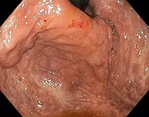
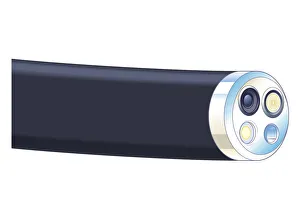
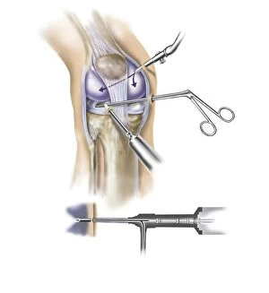
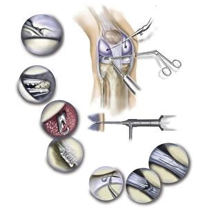
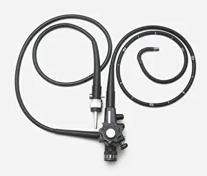
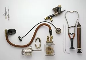
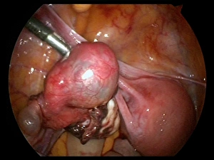
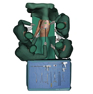
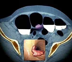
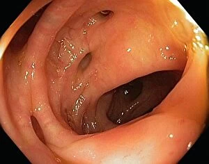
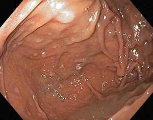
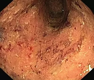
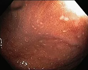
"Exploring the Inner World: Unveiling Medical Marvels with Endoscope" Journey into the Unknown: Witnessing an Ovarian Cyst through the Lens of an Endoscope (C017 / 6800) Delving Deeper: Peering Inside the Uterus with Endoscopic View (C017 / 6805) Unraveling Gastric Antral Vascular Ectasia: A Glimpse through Endoscopy (C016 / 8328) Navigating New Frontiers: Biomedical Illustration of Hysteroscopy Procedure using an Endoscope Unlocking Secrets within: Biomedical Illustration of Hysteroscopy Examination via Endoscope Illuminating Hidden Pathways: Exploring Nose and Throat with a Cross-Section Biomedical Illustration The Knee's Inner Story Revealed: Rigid Endoscopy Procedure Unveiled in a Cross-Section Biomedical Illustration Precision at its Finest: A Detailed Biomedical Illustration of the Tip of a Flexible Endoscope The Versatile Tool for Exploration: Discovering the Rigid Endoscope in all its Glory Laparoscopic Surgery Demystified: Abdominal Adventure Explored in a Cross-Section Biomedical Illustration Exploring Beneath the Surface - Arthroscopic Instruments Reveal Intricate Details of Human Knee Surgical Artistry at Work.