Fraud Blocker Skip to main content

Electron Microscope Collection

The electron microscope, a marvel of scientific innovation, has revolutionized our understanding of the microscopic world

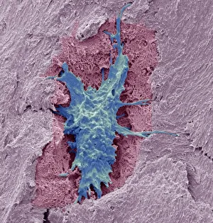
Background imageElectron Microscope Collection: Osteocyte bone cell, SEM C016 / 9025

Osteocyte bone cell, SEM C016 / 9025
Osteocyte bone cell. Coloured scanning electron micrograph (SEM) of an osteocyte bone cell (blue) surrounded by bone tissue (pink)

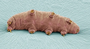
Background imageElectron Microscope Collection: Water bear, SEM C016 / 9084

Water bear, SEM C016 / 9084
Water bear (Echiniscus granulatus). Coloured scanning electron micrograph (SEM) showing the underside of a water bear, or tardigrade (phylum Tardigrada)

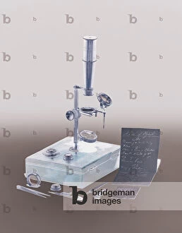
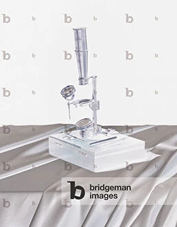
Background imageElectron Microscope Collection: A lacquered brass microscope belonging to the Darwin family, c.1826-30 (mixed media)

A lacquered brass microscope belonging to the Darwin family, c.1826-30 (mixed media)
8736176 A lacquered brass microscope belonging to the Darwin family, c.1826-30 (mixed media) by English School, (19th century); 13.7x10.7x3.7 cm; Private Collection; (add.info)

Background imageElectron Microscope Collection: Female American mathematician and astronomer at work, at the Paris Observatory, 1850

Female American mathematician and astronomer at work, at the Paris Observatory, 1850
3478832 Female American mathematician and astronomer at work, at the Paris Observatory, 1850; (add.info.: Miss Klumpke an American mathematician and astronomer at work on the Carte du Ciel)

Background imageElectron Microscope Collection: Title page of the first volume of Robert Brown's 'Science for All', 1850

Title page of the first volume of Robert Brown's 'Science for All', 1850
3331022 Title page of the first volume of Robert Brown's 'Science for All', 1850; (add.info.: Title page of the first volume of Robert Brown's 'Science for All')

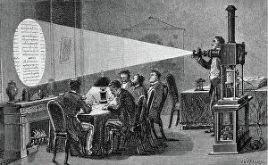
Background imageElectron Microscope Collection: Projection and enlargement of microscopic reports during the siege of Paris in 1871. (engraving)

Projection and enlargement of microscopic reports during the siege of Paris in 1871. (engraving)
PCT4256951 Projection and enlargement of microscopic reports during the siege of Paris in 1871. (engraving); (add.info.: The microphotography invented by Dagron allows the defenders of Paris to send)

Background imageElectron Microscope Collection: A lacquered brass microscope belonging to the Darwin family, c.1826-30 (mixed media)

A lacquered brass microscope belonging to the Darwin family, c.1826-30 (mixed media)
8736188 A lacquered brass microscope belonging to the Darwin family, c.1826-30 (mixed media) by English School, (19th century); 13.7x10.7x3.7 cm; Private Collection; (add.info)

Background imageElectron Microscope Collection: A lacquered brass microscope belonging to the Darwin family, c.1826-30 (mixed media)

A lacquered brass microscope belonging to the Darwin family, c.1826-30 (mixed media)
8736162 A lacquered brass microscope belonging to the Darwin family, c.1826-30 (mixed media) by English School, (19th century); 13.7x10.7x3.7 cm; Private Collection; (add.info)

Background imageElectron Microscope Collection: How man learns from the human eye (litho)

How man learns from the human eye (litho)
8676259 How man learns from the human eye (litho) by English School, (20th century); Private Collection; (add.info.: How man learns from the human eye. Inventions using lenses)

Background imageElectron Microscope Collection: Story of Sand: Sand under the microscope (colour litho)

Story of Sand: Sand under the microscope (colour litho)
8659632 Story of Sand: Sand under the microscope (colour litho) by English School, (20th century); Private Collection; (add.info.: Sand under the microscope)

Background imageElectron Microscope Collection: A lacquered brass microscope belonging to the Darwin family, c.1826-30 (mixed media)

A lacquered brass microscope belonging to the Darwin family, c.1826-30 (mixed media)
8736181 A lacquered brass microscope belonging to the Darwin family, c.1826-30 (mixed media) by English School, (19th century); 13.7x10.7x3.7 cm; Private Collection; (add.info)

Background imageElectron Microscope Collection: A lacquered brass microscope belonging to the Darwin family, c.1826-30 (mixed media)

A lacquered brass microscope belonging to the Darwin family, c.1826-30 (mixed media)
8736170 A lacquered brass microscope belonging to the Darwin family, c.1826-30 (mixed media) by English School, (19th century); 13.7x10.7x3.7 cm; Private Collection; (add.info)

Background imageElectron Microscope Collection: Microscope composed by Galileo Galilei and made by Giuseppe Campani, 1650-1700 (cardboard, wood)

Microscope composed by Galileo Galilei and made by Giuseppe Campani, 1650-1700 (cardboard, wood, metal and leather)
DEX5040791 Microscope composed by Galileo Galilei and made by Giuseppe Campani, 1650-1700 (cardboard, wood, metal and leather); Istituto e Museo di Storia della Scienza, Florence

Background imageElectron Microscope Collection: Microscope by the Zeiss company from Jena

Microscope by the Zeiss company from Jena
7277941 Microscope by the Zeiss company from Jena by Unknown Artist, (19th century); Private Collection; (add.info.: Microscope by the Zeiss company from Jena)

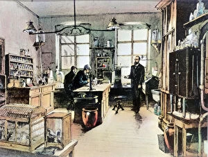
Background imageElectron Microscope Collection: Portrait of Robert Koch in his laboratory (engraving, 19th century)

Portrait of Robert Koch in his laboratory (engraving, 19th century)
LRI4670026 Portrait of Robert Koch in his laboratory (engraving, 19th century) by Unknown Artist, (19th century); Private Collection; (add.info)

Background imageElectron Microscope Collection: Study showing the form and structure of a Snowflake, c. 1902 (photo)

Study showing the form and structure of a Snowflake, c. 1902 (photo)
7252346 Study showing the form and structure of a Snowflake, c.1902 (photo) by Bentley, Wilson A. (1865-1931); Private Collection; (add.info.: Wilson Alwyn Bentley (1865 - 1931)

Background imageElectron Microscope Collection: Study showing the form and structure of a Snowflake, c. 1902 (photo)

Study showing the form and structure of a Snowflake, c. 1902 (photo)
7252352 Study showing the form and structure of a Snowflake, c.1902 (photo) by Bentley, Wilson A. (1865-1931); Private Collection; (add.info.: Wilson Alwyn Bentley (1865 - 1931)

Background imageElectron Microscope Collection: Study showing the form and structure of a Snowflake, c. 1902 (photo)

Study showing the form and structure of a Snowflake, c. 1902 (photo)
7252343 Study showing the form and structure of a Snowflake, c.1902 (photo) by Bentley, Wilson A. (1865-1931); Private Collection; (add.info.: Wilson Alwyn Bentley (1865 - 1931)

Background imageElectron Microscope Collection: Study showing the form and structure of a Snowflake, c. 1902 (photo)

Study showing the form and structure of a Snowflake, c. 1902 (photo)
7252353 Study showing the form and structure of a Snowflake, c.1902 (photo) by Bentley, Wilson A. (1865-1931); Private Collection; (add.info.: Wilson Alwyn Bentley (1865 - 1931)

Background imageElectron Microscope Collection: Study showing the form and structure of a Snowflake, c. 1902 (photo)

Study showing the form and structure of a Snowflake, c. 1902 (photo)
7252357 Study showing the form and structure of a Snowflake, c.1902 (photo) by Bentley, Wilson A. (1865-1931); Private Collection; (add.info.: Wilson Alwyn Bentley (1865 - 1931)

Background imageElectron Microscope Collection: Study of Snowflakes, c. 1903 (photo)

Study of Snowflakes, c. 1903 (photo)
7252363 Study of Snowflakes, c.1903 (photo) by Bentley, Wilson A. (1865-1931); Private Collection; (add.info.: Wilson Alwyn Bentley (1865 - 1931), also known as Snowflake Bentley

Background imageElectron Microscope Collection: Study showing the form and structure of a Snowflake, c. 1902 (photo)

Study showing the form and structure of a Snowflake, c. 1902 (photo)
7252351 Study showing the form and structure of a Snowflake, c.1902 (photo) by Bentley, Wilson A. (1865-1931); Private Collection; (add.info.: Wilson Alwyn Bentley (1865 - 1931)

Background imageElectron Microscope Collection: Study showing the form and structure of a Snowflake, c. 1902 (photo)

Study showing the form and structure of a Snowflake, c. 1902 (photo)
7252349 Study showing the form and structure of a Snowflake, c.1902 (photo) by Bentley, Wilson A. (1865-1931); Private Collection; (add.info.: Wilson Alwyn Bentley (1865 - 1931)

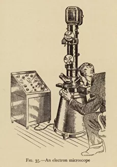
Background imageElectron Microscope Collection: Electron microscope (litho)

Electron microscope (litho)
7185637 Electron microscope (litho) by Adam, Victor (1801-66) (after); Private Collection; (add.info.: Electron microscope)

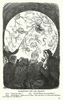
Background imageElectron Microscope Collection: Microscopy for the Million (litho)

Microscopy for the Million (litho)
7201243 Microscopy for the Million (litho) by Keene, Charles (1823-91); Private Collection; (add.info.: Microscopy for the million)

Background imageElectron Microscope Collection: Modern Times, the Electron Microscope (b / w photo)

Modern Times, the Electron Microscope (b / w photo)
3649875 Modern Times, the Electron Microscope (b/w photo) by English Photographer, (20th century); Private Collection; (add.info.: Modern Times, the Electron Microscope)

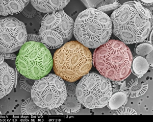
Background imageElectron Microscope Collection: Emiliania huxleyi coccolithophores

Emiliania huxleyi coccolithophores collected from a bloom in the SW Approaches to the English Channel in June 2004. Date: 2004

Background imageElectron Microscope Collection: Maggot head, SEM C014 / 1449

Maggot head, SEM C014 / 1449
Maggot head. Coloured scanning electron micrograph (SEM) of the head of a fly (order Diptera) larva, showing its mouthparts (centre). Magnification: x40 when printed at 10 centimetres wide

Background imageElectron Microscope Collection: Osteocyte bone cell, SEM C016 / 9026

Osteocyte bone cell, SEM C016 / 9026
Osteocyte bone cell. Coloured scanning electron micrograph (SEM) of an osteocyte bone cell (red) surrounded by bone tissue (grey)

Background imageElectron Microscope Collection: FE scanning electron microscopy C016 / 3821

FE scanning electron microscopy C016 / 3821
FE scanning electron microscopy. Researcher operating a field-emission scanning electron microscope (FE-SEM). This is a F JEOL 6320F FE-SEM

Background imageElectron Microscope Collection: Scanning transmission electron microscopy C016 / 3815

Scanning transmission electron microscopy C016 / 3815
Scanning transmission electron microscopy. Researcher using a scanning transmission electron microscope (STEM) to analyse the structure of solid-state materials

Background imageElectron Microscope Collection: Tabanidae. Horse Fly

Tabanidae. Horse Fly

Background imageElectron Microscope Collection: Midge larva, SEM C017 / 1912

Midge larva, SEM C017 / 1912
Midge larva. Coloured scanning electron micrograph (SEM) of a buzzer midge (Chironomus plumosus) larva. The buzzer midge, is a species of non-biting midge

Background imageElectron Microscope Collection: Spiderwort anther and pollen, SEM C017 / 1911

Spiderwort anther and pollen, SEM C017 / 1911
Spiderwort anther and pollen. Coloured scanning electron micrograph (SEM) of pollen grains (yellow) on hairs from the anther of a Spiderwort (Tradescantia sp.) flower

Background imageElectron Microscope Collection: Ladybird foot hairs, SEM C014 / 1447

Ladybird foot hairs, SEM C014 / 1447
Ladybird foot hairs. Coloured scanning electron micrograph (SEM) of the foot of a ladybird (family Coccinellidae), showing the numerous hairs that cover its surface

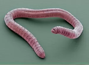
Background imageElectron Microscope Collection: Annelid worm, SEM C014 / 1428

Annelid worm, SEM C014 / 1428
Annelid worm. Coloured scanning electron micrograph (SEM) of an annelid (segmented) worm, showing small clumps of setae (bristles). Magnification: x20 when printed at 10 centimetres wide

Background imageElectron Microscope Collection: Phosphate crystals, SEM C014 / 1436

Phosphate crystals, SEM C014 / 1436
Phosphate crystals, coloured scanning electron micrograph (SEM). Magnification: x500 when printed at 10 centimetres wide

Background imageElectron Microscope Collection: Phosphate crystals, SEM C014 / 1435

Phosphate crystals, SEM C014 / 1435
Phosphate crystals, coloured scanning electron micrograph (SEM). Magnification: x500 when printed at 10 centimetres wide

Background imageElectron Microscope Collection: Annelid worm, SEM C014 / 1429

Annelid worm, SEM C014 / 1429
Annelid worm. Coloured scanning electron micrograph (SEM) of an annelid (segmented) worm, showing small clumps of setae (bristles). Magnification: x20 when printed at 10 centimetres wide

Background imageElectron Microscope Collection: Pseudomonas aeruginosa bacterium, SEM C016 / 9442

Pseudomonas aeruginosa bacterium, SEM C016 / 9442
Pseudomonas aeruginosa bacterium, coloured transmission electron micrograph (TEM). P. aeruginosa is a Gram-negative rod-shaped bacterium found in soil and water

Background imageElectron Microscope Collection: White blood cell antigen presentation C016 / 9058

White blood cell antigen presentation C016 / 9058
White blood cell antigen presentation. Coloured scanning electron micrograph (SEM) showing the interaction between a macrophage (yellow) and a T helper lymphocyte (Th cell, green)

Background imageElectron Microscope Collection: Water bear, SEM C016 / 9085

Water bear, SEM C016 / 9085
Water bear (Echiniscus granulatus). Coloured scanning electron micrograph (SEM) showing the underside of a water bear, or tardigrade (phylum Tardigrada)

Background imageElectron Microscope Collection: Microglial white blood cell, SEM C016 / 9115

Microglial white blood cell, SEM C016 / 9115
Microglial white blood cell. Coloured scanning electron micrograph (SEM) of a microglial cell. Microglia are phagocytic macrophage white blood cells that are the main form of immune system in

Background imageElectron Microscope Collection: Mycobacterium fortuitum bacteria C016 / 9424

Mycobacterium fortuitum bacteria C016 / 9424
Mycobacterium fortuitum bacteria, coloured scanning electron micrograph (SEM). These bacteria (blue) are a non-tuberculosis strain of Mycobacterium

Background imageElectron Microscope Collection: Lassa virus particles, TEM C016 / 9409

Lassa virus particles, TEM C016 / 9409
Lassa virus particles. Transmission electron micrograph (TEM) of Lassa virus particles (virions, blue) amongst cell debris. This Arenavirus is the cause of Lassa fever

Background imageElectron Microscope Collection: Dog tapeworm, SEM C016 / 9039

Dog tapeworm, SEM C016 / 9039
Dog tapeworm. Coloured scanning electron micrograph (SEM) of a dog tapeworm (Echinococcus granulosus). Tapeworms are parasitic flatworms that live in the digestive tract of their vertebrate host. E

Background imageElectron Microscope Collection: Orientia tsutsugamushi bacterium C016 / 9431

Orientia tsutsugamushi bacterium C016 / 9431
Orientia tsutsugamushi bacterium. Transmission electron micrograph (TEM) of a section through an Orientia tsutsugamushi bacterium. O

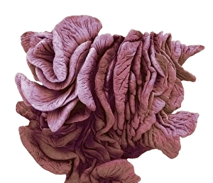
Background imageElectron Microscope Collection: Fimbriae of a fallopian tube, SEM C016 / 9047

Fimbriae of a fallopian tube, SEM C016 / 9047
Fimbriae of a fallopian tube, coloured scanning electron micrograph (SEM). Fimbriae are the folds at the opening (ostium) of a fallopian tube, next to an ovary


All Professionally Made to Order for Quick Shipping

The electron microscope, a marvel of scientific innovation, has revolutionized our understanding of the microscopic world. With its incredible magnification capabilities, it allows us to delve into the intricate details of various specimens and uncover hidden wonders. One such specimen is the osteocyte bone cell, captured in stunning detail by SEM C016 / 9025. This image reveals the complex structure and delicate nature of these cells that play a crucial role in maintaining healthy bones. In another captivating image (SEM C016 / 9084), we are introduced to the water bear, an extraordinary creature known for its ability to survive extreme conditions. Through the electron microscope's lens, we witness its unique anatomy and gain insight into how it adapts to different environments. Moving on from living organisms, we explore Emiliania huxleyi coccolithophores – tiny marine algae with intricately patterned shells. The electron microscope unveils their exquisite beauty and provides valuable information about their ecological significance, and is worth noting that this remarkable instrument owes its existence to companies like Zeiss from Jena who have dedicated themselves to pushing technological boundaries in microscopy. Their contributions have paved the way for groundbreaking discoveries across various scientific disciplines. Looking back through history, we encounter an engraving depicting Robert Koch in his laboratory during the 19th century. This iconic image reminds us of how far microscopy has come since then and highlights Koch's influential work in microbiology. Additionally, we are transported back over a century ago with photographs showcasing studies on snowflakes' form and structure around 1902-1903. These mesmerizing images capture nature's artistry at its finest while demonstrating how electron microscopes enable us to unravel even seemingly simple phenomena like snowflakes' intricate designs. The electron microscope continues to amaze scientists and enthusiasts alike with its ability to reveal hidden worlds beyond our naked eye's reach.