
Distal Collection
"Exploring the Distal Realm: From Kidney Tubules to Extinct Bones" Delve into the intricate world anatomy with a captivating array of images


All Professionally Made to Order for Quick Shipping
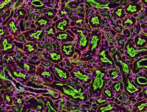
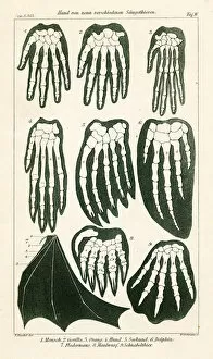
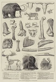
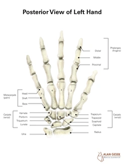
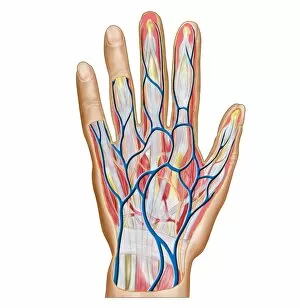
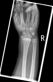
"Exploring the Distal Realm: From Kidney Tubules to Extinct Bones" Delve into the intricate world anatomy with a captivating array of images. Starting with kidney tubules in section, we witness the complexity and functionality of these vital organs. Moving on, a fascinating study from 1898 showcases a colorful lithograph comparing hand bones across nine different mammals, highlighting their unique adaptations. Unearthing ancient secrets, an engraving reveals the bones of extinct animals discovered at Charing-Cross. The posterior view of a left hand provides detailed labels that unravel its anatomical wonders. Another image presents the conceptual beauty of human hand bones, showcasing their elegance and structural brilliance. Continuing our exploration, we delve deeper into the backside anatomy of the human hand - every tendon and muscle intricately depicted for us to marvel at. A three-dimensional view unveils the female skeletal system in all its glory - an awe-inspiring testament to nature's design. Returning to hands once more, we encounter another conceptual image emphasizing their bone structure as if it were art itself. But there is more than meets the eye; a human hand comes alive when accompanied by its nervous system, lymphatic system, and circulatory system - an interconnected masterpiece. Finally, shifting focus towards reproduction biology, we are presented with an intimate glimpse into male testis anatomy. This comprehensive collection invites us to appreciate both our shared heritage with other mammals and our own remarkable uniqueness as humans. In this captivating journey through various aspects anatomy imagery – from kidneys to hands and beyond – one cannot help but be awestruck by nature's incredible designs and complexities that lie within each living being.