
Dicot Collection (page 74)
"Dicot: A Journey through Nature's Diversity" Explore the fascinating world of dicots, a diverse group of plants that includes some truly remarkable species


All Professionally Made to Order for Quick Shipping
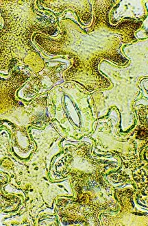
"Dicot: A Journey through Nature's Diversity" Explore the fascinating world of dicots, a diverse group of plants that includes some truly remarkable species. From the rare and elusive Franklinia alatamaha, also known as Franklinia, to the vibrant Hydrangea hortensis or French hydrangea, each dicot offers its own unique beauty. Find solace under the graceful branches of a Weeping Willow or marvel at the intricate patterns on insects found in Surinam. Discover the humble Gossypium barbadense, commonly known as cotton plant, which has played a significant role in shaping our history. Immerse yourself in the enchanting Scottish Pine Forest or wander through Lineover Wood in Gloucestershire UK, where ancient Beech trees (Fagus sylvatica) stand tall with wisdom accumulated over centuries. Indulge your senses with Durio zibethinus - durian fruit's distinctive aroma and taste. Experience the serene beauty of heathland landscapes that harbor an array of unique flora and fauna. Savor Orangier des Gcnes or Arancio di Genova oranges for their exquisite flavor and fragrance. Uncover the secrets hidden within Myristica sp. , better known as nutmeg - a spice cherished for its warm essence. Lastly, witness nature's ingenuity with Cephalotus follicularis - Australian pitcher plant - showcasing its carnivorous adaptations to thrive in nutrient-poor environments. Dicots offer us glimpses into nature's vast creativity and resilience. They remind us of our interconnectedness with all living beings and inspire us to appreciate Earth's incredible biodiversity. Let these hints guide you on an awe-inspiring journey through this captivating realm.