Fraud Blocker Skip to main content

Diagnosis Collection (page 4)

"Unlocking the Secrets: The Power of Diagnosis" A seahorse's hidden world revealed through an X-ray, reminding us that diagnosis goes beyond human health

Background imageDiagnosis Collection: Bladder stones, ultrasound scan C017 / 7974

Bladder stones, ultrasound scan C017 / 7974
Bladder stones. Ultrasound scan of the bladder of a 58 year old male showing bladder stones (bright white). Bladder stones (cystoliths)

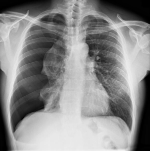
Background imageDiagnosis Collection: Pneumothorax, X-ray C017 / 7812

Pneumothorax, X-ray C017 / 7812
Pneumothorax. X-ray of the chest of a patient with a pneumothorax, or collapsed lung. The right lung (left on X-ray) has collapsed due to a build-up of air (solid grey)

Background imageDiagnosis Collection: Hydropneumothorax, X-ray C017 / 7810

Hydropneumothorax, X-ray C017 / 7810
Hydropneumothorax. X-ray of the chest of a female patient with a hydropneumothorax of the right lung (seen at left). This is a condition where air and fluid accumulate in the spaces around the lungs

Background imageDiagnosis Collection: Endometrial polyp, ultrasound scan C017 / 7800

Endometrial polyp, ultrasound scan C017 / 7800
Endometrial polyp. Ultrasound scan of the uterus (womb) of a 42 year old female patient with a polyp (lower centre) in the endometrium, the lining of the womb

Background imageDiagnosis Collection: Polycystic kidney, ultrasound scan C017 / 7746

Polycystic kidney, ultrasound scan C017 / 7746
Polycystic kidney. Ultrasound scan of the kidney of a 42 year old female showing numerous fluid-filled cysts (dark areas)

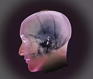
Background imageDiagnosis Collection: Deep brain stimulation, X-ray

Deep brain stimulation, X-ray
Image Xrays Crane (Sur Projection 3D Modèle) En Incidence De Profil Chez Une Patiente De 54 Ans, Parkinsonienne Traitee Sous Protocole De Neurochirurgie; Visualisation Des Electrodes De

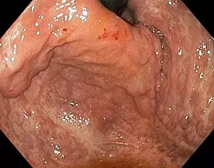
Background imageDiagnosis Collection: Severe gastritis, endoscope view C016 / 3898

Severe gastritis, endoscope view C016 / 3898
Severe gastritis, endoscope view. Gastritis is inflammation of the stomach lining. It causes pain on eating or drinking, and may cause nausea and vomiting

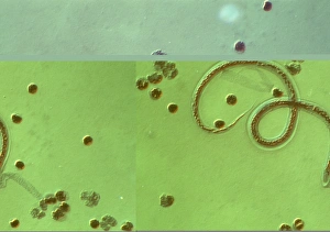
Background imageDiagnosis Collection: Wuchereria bancrofti parasite

Wuchereria bancrofti parasite
Wuchereria bancrofti. Light micrograph of the microfilaria larval stage of the parasitic worm Wuchereria bancrofti, which causes filariasis in humans. W

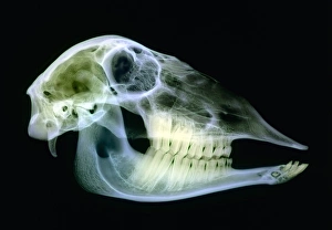
Background imageDiagnosis Collection: Sheep skull

Sheep skull. Coloured X-ray of a sagittal section through a sheeps (Ovis aries) skull. Sheep have 32 permanent teeth; 12 molars and eight incisors on the bottom jaw and 12 molars on the top jaw

Background imageDiagnosis Collection: Naked womans body with aura, artwork

Naked womans body with aura, artwork
Naked woman. Computer artwork of a naked woman with an aura around her body. Aura therapists consider the aura to be the natural life force surrounding a person

Background imageDiagnosis Collection: Fractured foot, coloured X-ray

Fractured foot, coloured X-ray
Fractured foot. Coloured X-rays of a fractured fifth metatarsal (to the right of each image). The fracture is seen as a small black crack

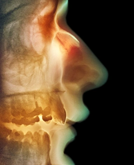
Background imageDiagnosis Collection: Broken nose, X-ray

Broken nose, X-ray
Broken nose. Coloured X-ray of a side view of a mans head showing a broken nose (upper centre). The fracture of the nasal bone appears as a thin line

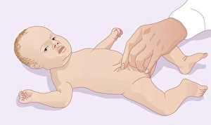
Background imageDiagnosis Collection: Skin pinch test for dehydration, artwork

Skin pinch test for dehydration, artwork
Skin pinch test for dehydration. Computer artwork of a doctor pinching a babys skin to test for dehydration. The longer the skin takes to return after pinching, the more severe the dehydration

Background imageDiagnosis Collection: Lung abscess, X-ray

Lung abscess, X-ray
Lung abscess. Coloured X-ray showing an abscess (dense coloured area, upper right) in the upper lobe of a patients left lung (right). An abscess is an accumulation of puss within a cavity in tissue

Background imageDiagnosis Collection: Multiple sclerosis, MRI scan

Multiple sclerosis, MRI scan

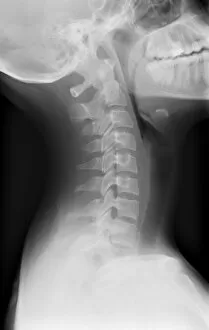
Background imageDiagnosis Collection: Healthy spine of the neck, X-ray

Healthy spine of the neck, X-ray
X-ray (side view) of the neck of an adult patient, showing healthy cervical vertebrae (spine bones) of the neck. The skull (at top) is tilted back, with the shoulders at lower frame

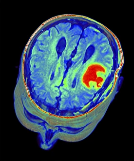
Background imageDiagnosis Collection: Brain tumour, 3D-MRI scan

Brain tumour, 3D-MRI scan
Brain tumour. Coloured three-dimensional model of a male head, made up from numerous magnetic resonance imaging (MRI) scans

Background imageDiagnosis Collection: Native Doctor - Sierra Leone

Native Doctor - Sierra Leone. Checking a diagnosis by the means of the placement of a set of small stones of seeds

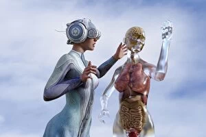
Background imageDiagnosis Collection: android, ar, augmented reality, body, brain, cloud, colon, color image, concept, copy space

android, ar, augmented reality, body, brain, cloud, colon, color image, concept, copy space
Woman viewing organs of transparent woman in virtual reality

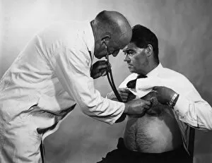
Background imageDiagnosis Collection: Checking Heartbeat

Checking Heartbeat
A doctor listens to a patients heart Date: 1960s

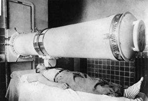
Background imageDiagnosis Collection: Cancer X-Ray Machine

Cancer X-Ray Machine
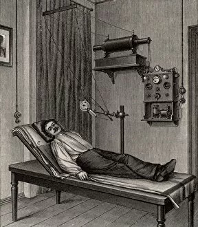
A huge new German x-ray lamp, for cancer treatment, which casts a very long localised beam on the affected area, without touching the rest of the patients body. Date: 1933

Background imageDiagnosis Collection: Mongrel puppy identified by the vet

Mongrel puppy identified by the vet -- he is a mongrel who has already reached his full size. Date: 1914

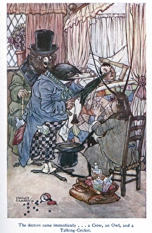
Background imageDiagnosis Collection: Pinocchio -- ill in bed

Pinocchio -- ill in bed
Pinocchio -- The doctors came immediately... a Crow, an Owl, and a Talking Cricket. Date: 1911

Background imageDiagnosis Collection: Richard Powell

Richard Powell
RICHARD POWELL Doctor at St Bartholomews hospital, London, who was responsible for many valuable innovations in diagnosis and treatment. Date: 1767 - 1834

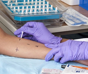
Background imageDiagnosis Collection: A person receiving a skin prick allergy test

A person receiving a skin prick allergy test
A person receiving a skin prick test (SPT). An SPT is performed by using a needle to place a tiny amount of liquid allergen just under the skin of the forearm

Background imageDiagnosis Collection: The Doctors Visit (oil on canvas)

The Doctors Visit (oil on canvas)
XIR223847 The Doctors Visit (oil on canvas) by Dullaert, Heiman (1636-1684); Groningen, The Netherlands; Dutch, out of copyright

Background imageDiagnosis Collection: The Consultation, c. 1655 (oil on canvas)

The Consultation, c. 1655 (oil on canvas)
XIR223846 The Consultation, c.1655 (oil on canvas) by Brekelenkam, Quiringh Gerritsz. van (c.1620-68); 57x52 cm; Louvre, Paris, France; Dutch, out of copyright

Background imageDiagnosis Collection: The Urine Examination (coloured engraving)

The Urine Examination (coloured engraving)
XIR223854 The Urine Examination (coloured engraving) by German School, (18th century); Museo Nazionale Dell Arte Sanitaria, Rome, Italy; German, out of copyright

Background imageDiagnosis Collection: The Patient and the Doctor (oil on panel)

The Patient and the Doctor (oil on panel)
XIR173150 The Patient and the Doctor (oil on panel) by Steen, Jan Havicksz. (1625/26-79); Narodni Galerie, Prague, Czech Republic; Dutch, out of copyright

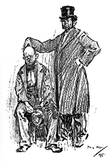
Background imageDiagnosis Collection: Unhelpful Phrenological assessment of a Park Loafer

Unhelpful Phrenological assessment of a Park Loafer. Phrenologist (to Park Loafer) - " Now, you re full o Nerves, you are

Background imageDiagnosis Collection: Rene Theophile Hyacinthe Laennec (1781-1826), French physician who invented the Stethoscope

Rene Theophile Hyacinthe Laennec (1781-1826), French physician who invented the Stethoscope. (1889). Laennec is shown listening to a patients chest at the Necker Hospital, Paris

Background imageDiagnosis Collection: Physician examining a specimen of patients urine (16th-17th century) 19th century

Physician examining a specimen of patients urine (16th-17th century) 19th century chromolithograph

Background imageDiagnosis Collection: Avicenna (Ibn Sina) 980-1037, Arab physician and philosopher. An interpreter of Aristotle to Islam

Avicenna (Ibn Sina) 980-1037, Arab physician and philosopher. An interpreter of Aristotle to Islam: compiled encyclopaedia of medical knowledge

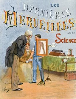
Background imageDiagnosis Collection: X-ray being taken of a boys shoulder. Title page of Les dernieres merveilles

X-ray being taken of a boys shoulder. Title page of Les dernieres merveilles de la science (The Latest Marvels of Science), Paris, c1895. Electricity Battery Acid Ruhmkorff Coil Medicine Diagnosis

Background imageDiagnosis Collection: La Pharmacie Rustique, c1775 (The Country Pharmacy). The composition shows a

La Pharmacie Rustique, c1775 (The Country Pharmacy). The composition shows a visit to a country chemist. Illustrated with the interior of Michael Schuppachs pharmacy in Basel, Switzerland

Background imageDiagnosis Collection: X-ray being taken of a patients thorax. The X-ray pate is beneath the patient at C

X-ray being taken of a patients thorax. The X-ray pate is beneath the patient at C. From Die Naturkrafte by M Wilhelm Meyer (Leipzig, 1903)

Background imageDiagnosis Collection: Testing strip being dipped into a urine sample

Testing strip being dipped into a urine sample

Background imageDiagnosis Collection: Broken elbow, X-ray C017 / 7266

Broken elbow, X-ray C017 / 7266
Broken elbow. X-ray of the elbow of a 4 year old female with a displaced supracondylar fracture. The fracture is at the base of the humerus, the upper arm bone (upper frame)

Background imageDiagnosis Collection: Arthritic hand, X-ray C017 / 7171

Arthritic hand, X-ray C017 / 7171
Arthritic hand. X-ray of the hand and wrist of a 56 year old patient with rheumatoid arthritis. Rheumatoid arthritis is caused by the immune system attacking the bodys own tissues

Background imageDiagnosis Collection: Secondary bone cancer, X-ray C017 / 7162

Secondary bone cancer, X-ray C017 / 7162
Secondary bone cancer. X-ray of the spine of a 40 year old female with metastatic (secondary) bone cancer (bright white areas). The cancer has metastasised (spread) from a primary cancer

Background imageDiagnosis Collection: Telemedicine, conceptual image C017 / 7605

Telemedicine, conceptual image C017 / 7605
MODEL RELEASED. Telemedicine, conceptual image

Background imageDiagnosis Collection: Telemedicine, conceptual image C017 / 7592

Telemedicine, conceptual image C017 / 7592
MODEL RELEASED. Telemedicine, conceptual image

Background imageDiagnosis Collection: Broken elbow, X-ray C017 / 7583

Broken elbow, X-ray C017 / 7583
Broken elbow. X-ray of the arm of a 55 year old male patient with fractures to the elbow and radius (upper lower arm bone)

Background imageDiagnosis Collection: Telemedicine, conceptual image C017 / 7590

Telemedicine, conceptual image C017 / 7590
Telemedicine, conceptual image

Background imageDiagnosis Collection: Telemedicine, conceptual image C017 / 7607

Telemedicine, conceptual image C017 / 7607
Telemedicine, conceptual image

Background imageDiagnosis Collection: Scoliosis of the spine, X-ray C017 / 7167

Scoliosis of the spine, X-ray C017 / 7167
Scoliosis. X-ray of the spine of a 71 year old female patient with severe scoliosis (sideways curvature). This condition usually starts in infancy or adolescence, and is most often seen in girls

Background imageDiagnosis Collection: Telemedicine, conceptual image C017 / 7601

Telemedicine, conceptual image C017 / 7601
Telemedicine, conceptual image

Background imageDiagnosis Collection: Telemedicine, conceptual image C017 / 7603

Telemedicine, conceptual image C017 / 7603
MODEL RELEASED. Telemedicine, conceptual image


All Professionally Made to Order for Quick Shipping

"Unlocking the Secrets: The Power of Diagnosis" A seahorse's hidden world revealed through an X-ray, reminding us that diagnosis goes beyond human health. Unveiling the spectrum: a color blindness test helping us understand how our eyes perceive the world differently. Eyesight test chart guiding us towards clarity and precision in diagnosing visual impairments. Peering into fractures: a broken wrist bone captured on X-ray, aiding doctors in accurate diagnosis and treatment plans. Exploring beneath the surface: a panoramic dental X-ray exposing hidden oral issues for comprehensive diagnoses. Empowering individuals with knowledge: home cholesterol test kits providing early detection for proactive health management. Saving lives through swift intervention: Pneumothorax treatment guided by an X-ray to restore lung function effectively. Knees tell stories too: a normal knee showcased on an X-ray, enabling precise diagnoses of joint conditions and injuries. Delving deeper into mysteries within: full body scans via MRI revealing intricate details for accurate medical assessments. Artistry meets science as ECGs portray the rhythm of a healthy heart, serving as diagnostic masterpieces in healthcare settings. Battling tuberculosis head-on with an X-ray, unveiling its presence so prompt treatments can be initiated promptly