Fraud Blocker Skip to main content

Dendrites Collection (page 4)

"Dendrites: The Intricate Extensions of Nerve Cells Unveiled" Exploring the Cerebellum Tissue

Background imageDendrites Collection: Nerve cell culture, SEM

Nerve cell culture, SEM
Nerve cell culture. Coloured scanning electron micrograph (SEM) of new growth from a cultured sample from a spinal cord. The numerous branching strands are neurites

Background imageDendrites Collection: Cerebral cortex nerve cells

Cerebral cortex nerve cells

Background imageDendrites Collection: Purkinje nerve cell, SEM

Purkinje nerve cell, SEM

Background imageDendrites Collection: Purkinje nerve cell

Purkinje nerve cell

Background imageDendrites Collection: Purkinje nerve cells, SEM

Purkinje nerve cells, SEM
Purkinje nerve cells. Scanning electron micrograph (SEM) of two Purkinje nerve cells from the cerebellum of the brain. The cells comprise a flask-shaped cell body

Background imageDendrites Collection: Brain power

Brain power. Conceptual computer artwork of a Rubiks cube with neurological images on its surfaces. At top is a coloured magnetic resonance imaging (MRI) scan of a section through the head

Background imageDendrites Collection: Pain, conceptual computer artwork

Pain, conceptual computer artwork
Pain. Conceptual computer artwork of pain, where the site of pain is represented by a ball of barbed wire. Connected to this are the free nerve endings, which respond to mechanical

Background imageDendrites Collection: DNA and nerve cell, computer artwork

DNA and nerve cell, computer artwork
DNA (Deoxyribonucleic acid) strand and a nerve cell, computer artwork

Background imageDendrites Collection: Cerebellum structure, light micrograph

Cerebellum structure, light micrograph
Cerebellum structure. Coloured light micrograph of a section through the highly-folded cerebellum of the brain. The cerebellum comprises three main layers

Background imageDendrites Collection: Purkinje cells, light micrograph

Purkinje cells, light micrograph
Purkinje cells. Light micrograph of a section through the cerebellum, which has been treated with silver stains, showing pukinje cells (dark blue) and their dendritic processes

Background imageDendrites Collection: Nerve cell, computer artwork

Nerve cell, computer artwork
Nerve cell. Computer artwork of a nerve cell, or neuron

Background imageDendrites Collection: Pyramidal neurons, light micrograph

Pyramidal neurons, light micrograph with Nissl staining. This sample, from a human brain, shows two pyramidal neurons. Pyramidal neurons (nerve cells)

Background imageDendrites Collection: Purkinje neurons, light micrograph

Purkinje neurons, light micrograph

Background imageDendrites Collection: Nerve cells, artwork

Nerve cells, artwork
Neuron, nerve cell, firing, computer artwork

Background imageDendrites Collection: Muscle motor neurones, light micrograph

Muscle motor neurones, light micrograph
Muscle motor neurones. Light micrograph of dendrites (axons, black) from motor nerve cells (motor neurones) in muscle tissue (pink-purple strands)

Background imageDendrites Collection: Motor neurones, light micrograph

Motor neurones, light micrograph
Motor neurones. Light micrograph of motor nerve cells (motor neurones) and their support cells. The cell bodies of the neurones (large deep purple areas) have a central nucleus

Background imageDendrites Collection: Nerve synapse, artwork

Nerve synapse, artwork
Nerve synapse. Computer artwork of of a junction, or synapse, between two nerve cells (neurons). As the electrical signal reaches the presynaptic end of a neuron it triggers the release of

Background imageDendrites Collection: Nerve fibre, artwork

Nerve fibre, artwork
Nerve fibre, computer artwork

Background imageDendrites Collection: Nerve cell anatomy, artwork

Nerve cell anatomy, artwork
Nerve cell anatomy. Computer artwork showing the organelles in a neurone (nerve cell). At centre is nucleus (grey) and nucleolus (black)

Background imageDendrites Collection: Nerve cell and axon, diagram

Nerve cell and axon, diagram
Nerve cell and axon. Diagram showing how the input to a nerve cell (neuron) through dendrites (shown by arrows at upper left) is transmitted (yellow arrow) along the nerve cells axon (across bottom)

Background imageDendrites Collection: Parkinsons disease, artwork

Parkinsons disease, artwork
Parkinsons disease. Computer artwork of neurons (nerve cells, pink) containing Lewy bodies (green). Lewy bodies, which are deposits of protein

Background imageDendrites Collection: Mirror neurons, conceptual image

Mirror neurons, conceptual image
Mirror neurons, conceptual computer artwork. Nerve cells, or neurons, and a human eye reflected on a shiny surface representing a mirror neuron cell. Mirror neurons are found in the brain

Background imageDendrites Collection: Taste bud anatomy, diagram

Taste bud anatomy, diagram
Taste bud anatomy. Diagram of the anatomical structure of a taste bud on a tongue. The surface of the tongue is at bottom, with the tastebud (rounded structure) at centre

Background imageDendrites Collection: Neural network, artwork

Neural network, artwork
Neural network, computer artwork


All Professionally Made to Order for Quick Shipping

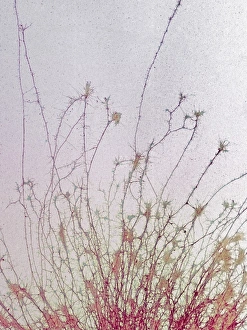
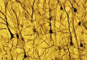
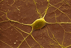
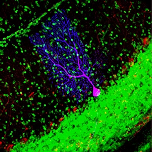
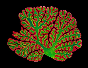
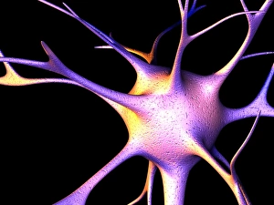
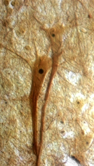
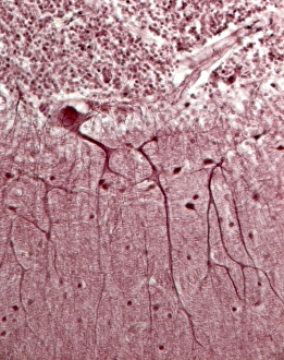
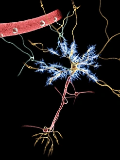
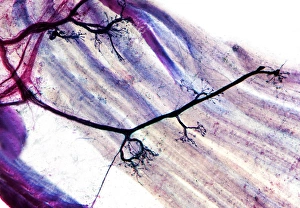
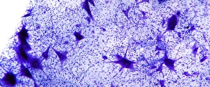
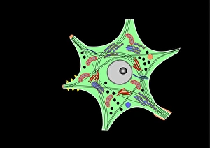
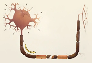
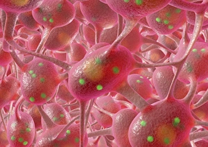
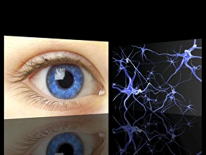
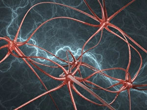
"Dendrites: The Intricate Extensions of Nerve Cells Unveiled" Exploring the Cerebellum Tissue: A mesmerizing light micrograph reveals the intricate network in this vital brain region. Unlocking Secrets at a Microscopic Level: Witness the stunning SEM image showcasing nerve cells and their elaborate dendritic branches, unraveling the mysteries of neural communication. Purkinje Nerve Cells in Action: Behold the captivating beauty of Purkinje nerve cells within the cerebellum, with their remarkable dendritic arborizations enabling precise motor control. Delving into Neuronal Complexity: Dive deep into this awe-inspiring artwork depicting dendritic cells, highlighting their crucial role in information processing and transmission within our nervous system. Unveiling Nature's Masterpiece: Another glimpse into Purkinje nerve cells' extraordinary architecture within the cerebellum, where countless dendrites intertwine to orchestrate seamless coordination. Illuminating Cerebellar Structure: This striking light micrograph showcases the complex organization within cerebellar tissue, emphasizing its significance for balance and motor functions. Insight into Motor Neurons' World: Witness how these vibrant light micrographs capture motor neurons' intricate web-like structures adorned with numerous branching dendrites responsible for muscle control. Marvels Within Cerebellum Tissue Revealed Again. Feast your eyes on yet another breathtaking light micrograph exposing intricately entangled dendrite networks that make up this critical brain region. Embracing Diversity in Neural Connections: Discover an array of nerve cells through abstract artwork, symbolizing diverse shapes and sizes of their fascinatingly branched dendritic extensions.