Fraud Blocker Skip to main content

Cytoskeleton Collection (page 3)

The cytoskeleton, a complex network of protein structures, plays a vital role in maintaining the shape and structure of cells

Background imageCytoskeleton Collection: Cells, SEM

Cells, SEM
Cell cytoskeleton. Coloured scanning electron micrograph (SEM) of the cytoskeleton (blue) and nuclei (green) of cultured cells

Background imageCytoskeleton Collection: Cytokinesis, artwork

Cytokinesis, artwork
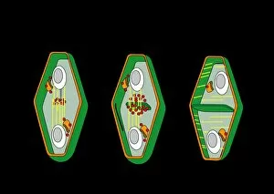
Cytokinesis. Artwork showing the stage of cell division that involves the splitting of the cell cytoplasm between two daughter cells

Background imageCytoskeleton Collection: Cytoskeleton and membrane, diagram

Cytoskeleton and membrane, diagram
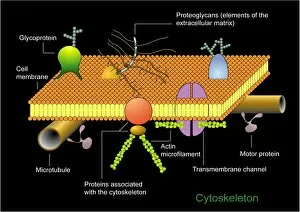
Cytoskeleton and membrane. Diagram showing the various structures associated with the cytoskeleton, the protein scaffolding found within cells

Background imageCytoskeleton Collection: Cytokinesis, diagram

Cytokinesis, diagram
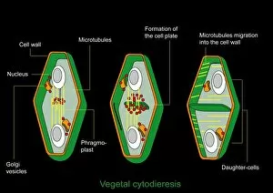
Cytokinesis. Diagram showing the stage of cell division that involves the splitting of the cell cytoplasm between two daughter cells

Background imageCytoskeleton Collection: Strychnine seed tissue, light micrograph

Strychnine seed tissue, light micrograph
Strychnine seed tissue. Light micrograph of a section through a seed of the strychnine plant (Strychnos nux vomica), showing the cellular bridges (plasmodesma)

Background imageCytoskeleton Collection: Cilium and flagellum structure, artwork

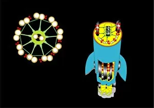
Cilium and flagellum structure, artwork. Cilia and flagella (collectively known as undulipodia) are hair-like protrusions from a cell membrane

Background imageCytoskeleton Collection: Hippocampal neuron fluorescent micrograph

Hippocampal neuron fluorescent micrograph
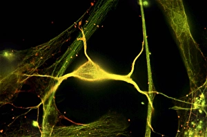
Hippocampal neuron. Fluorescent micrograph of a neuron (nerve cell, centre) from the hippocampus, surrounded by glial cells (support cells)

Background imageCytoskeleton Collection: Cell structure, fluorescent micrograph

Cell structure, fluorescent micrograph
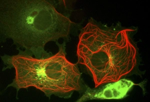
Cell structure. Fluorescent light micrograph of cultured cells from a cell line derived from African green monkey kidney cells. Microtubules, part of the cells cytoskeleton, are red

Background imageCytoskeleton Collection: Cytoskeleton components, artwork

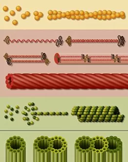
Cytoskeleton components, artwork. Three main components of the cytoskeleton, the internal support structure of a cell, are shown here

Background imageCytoskeleton Collection: Cytoskeleton components, diagram

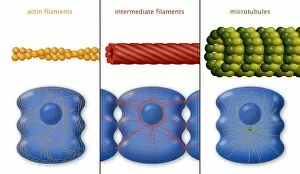
Cytoskeleton components, diagram. The cytoskeleton is the internal support structure of a cell, composed of filaments of various diameters in nanometres (nm)

Background imageCytoskeleton Collection: Animal cell processes, artwork

Animal cell processes, artwork
Animal cell processes. Cutaway artwork showing the structures inside an animal cell and four different processes that take place inside it or on its membrane (all marked by magnifying glasses)

Background imageCytoskeleton Collection: Animal cell structure, artwork

Animal cell structure, artwork
Animal cell structure. Artwork showing the internal structure of an animal cell. Inside the cell, the cellular structures (organelles) include the Golgi apparatus (green)

Background imageCytoskeleton Collection: Lung cells, fluorescent micrograph

Lung cells, fluorescent micrograph
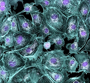
Lung cells. Immunofluorescence light micrograph of pulmonary endothelial cells. Endothelial cells are specialized epithelial cells that line the inner surface of blood vessels

Background imageCytoskeleton Collection: Microtubules, artwork

Microtubules, artwork
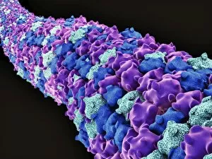
Microtubules, 3D computer artwork. Microtubules are polymers of the protein tubulin. They are a component of the cytoskeleton, which maintains a cells shape

Background imageCytoskeleton Collection: Protozoan tentacle, TEM

Protozoan tentacle, TEM
Protozoan tentacle. Coloured transmission electron micrograph (TEM) of a cross-section through a tentacle of a Dendrocometes sp. protozoan showing the microtubule (purple circles) structure

Background imageCytoskeleton Collection: Cytoskeleton, TEM

Cytoskeleton, TEM
Cytoskeleton. Coloured transmission electron micrograph (TEM) of the cytoskeleton of a human skin cell. The cell nucleus is at centre right

Background imageCytoskeleton Collection: Protein, microtubules and cell, artwork

Protein, microtubules and cell, artwork
3D computer artwork of a protein attached to microtubules, transporting vesicles and other important parts of the cell. Microtubules are polymers of the protein tubulin

Background imageCytoskeleton Collection: Cytoskeleton, SEM

Cytoskeleton, SEM
Cytoskeleton. Coloured scanning electron micrograph (SEM) of the cytoskeleton of a human skin cell. The cell nucleus is oval. The rest of the cells contents have been biochemically extracted

Background imageCytoskeleton Collection: Keratinocyte skin cells, light micrograph

Keratinocyte skin cells, light micrograph
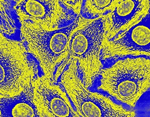
Keratinocyte skin cells. Fluorescent light micrograph of the cytoskeleton of human keratinocyte skin cells. Cell nuclei are oval. The rest of the cells contents have been biochemically extracted

Background imageCytoskeleton Collection: Movement of Listeria bacteria

Movement of Listeria bacteria
Movement of Listeria sp. bacteria. Immunofluorescence deconvolution micrograph showing the comet-like tails that help to propel Listeria sp. bacteria from cell to cell during an infection

Background imageCytoskeleton Collection: Actin tail, fluorescent micrograph

Actin tail, fluorescent micrograph
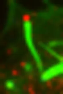
Actin tail. Immunofluorescence deconvolution micrograph of a close-up of an actin filament (green) in a cell infected with vaccinia virus particles

Background imageCytoskeleton Collection: Vaccinia virus infected cell

Vaccinia virus infected cell. Immunofluorescence deconvolution micrograph of a cell infected with vaccinia virus particles. Host and viral DNA (deoxyribonucleic acid) is blue

Background imageCytoskeleton Collection: E. coli infection mechanism

E. coli infection mechanism. Immunofluorescence deconvolution micrograph of Escherichia coli bacteria (centre). Bacterial DNA (deoxyribonucleic acid) is blue

Background imageCytoskeleton Collection: Fibroblast cells, fluorescent micrograph

Fibroblast cells, fluorescent micrograph
Fibroblast cells. Immunofluorescence deconvolution micrograph of fibroblast cells. The cell nuclei, which contain the cells genetic information, are blue

Background imageCytoskeleton Collection: Neural stem cells in culture

Neural stem cells in culture
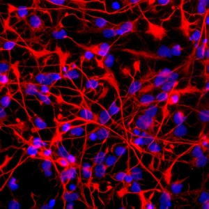
Neural stem cell in culture, fluorescent light micrograph. The stem cells have been dyed for nestin (red), an intermediate filament (IF) protein, and the nuclei are dyed blue

Background imageCytoskeleton Collection: Glial cells, SEM

Glial cells, SEM
Glial cells. Coloured scanning electron micrograph (SEM) of a glial cell (centre). Glial cells are nervous system cells that provide structural support and protection for neurons (nerve cells)

Background imageCytoskeleton Collection: Cytoskeleton, confocal light micrograph

Cytoskeleton, confocal light micrograph. Tubulin, the protein that makes up microtubules, is blue. Microtubules are part of the cytoskeleton, which maintains the cells shape

Background imageCytoskeleton Collection: Nerve cells and glial cells, SEM

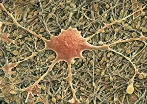
Nerve cells and glial cells, SEM
Nerve cells and glial cells, coloured scanning electron micrograph (SEM). The nerve cells have small cell bodies (orange) and fine extensions called axons and dendrites (brown)

Background imageCytoskeleton Collection: Glial cell, SEM

Glial cell, SEM
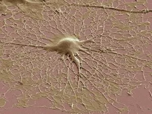
Glial cell. Coloured scanning electron micrograph (SEM) of a cultured glial cell (centre). Glial cells are nervous system cells that provide structural support and protection for neurons (nerve cells)

Background imageCytoskeleton Collection: Yeast cell, electron tomogram

Yeast cell, electron tomogram image. Yeast cell, Schizosaccharomyces pombe, created using a 3-D electron microscope. This involves firing beams of electrons from many different angles to create


All Professionally Made to Order for Quick Shipping

The cytoskeleton, a complex network of protein structures, plays a vital role in maintaining the shape and structure of cells. In budding yeast cells, it ensures proper cell division and growth. Nerve and glial cells rely on the cytoskeleton for their intricate connections, as seen in stunning light micrographs. Glial cells also exhibit their unique structure under confocal light microscopy. HeLa cells, another type of human cell line, reveal the intricate web-like nature of the cytoskeleton when observed through a light microscope. This structural framework supports various cellular processes within HeLa cells. Protozoa, fascinating single-celled organisms that scavenge for particles or absorb nutrients from their environment, also possess a cytoskeletal system to maintain their shape and enable movement. The microscopic view showcases this essential feature. Artwork depicting proteins interacting with microtubules highlights how these components contribute to overall cell function. Microtubules serve as highways for transporting materials within the cell. Conceptual images further illustrate the significance of the cytoskeleton across different organisms. Radiolarians showcase an intricate skeletal frame that provides support and protection while allowing them to move gracefully through water. A conceptual image of a plant cell emphasizes its various components supported by the cytoskeleton. Whether it's shaping budding yeast cells or enabling movement in protozoa or radiolarians, the cytoskeleton is an indispensable component responsible for maintaining cellular architecture and facilitating crucial biological processes across diverse organisms.