
Cut Away Collection (page 5)
"Unveiling the Inner Workings: Exploring the Fascinating World of Cutaways" Step into a world where secrets are revealed and hidden depths are exposed


All Professionally Made to Order for Quick Shipping
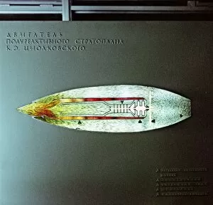
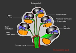
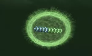
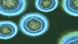
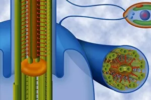
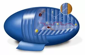
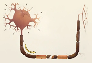
"Unveiling the Inner Workings: Exploring the Fascinating World of Cutaways" Step into a world where secrets are revealed and hidden depths are exposed. From vintage travel posters to wartime weaponry, from majestic architecture to intricate scientific devices, cutaway illustrations offer us a unique glimpse into the inner workings of various subjects. Immerse yourself in the golden age of aviation with an Imperial Airways poster showcasing four types of planes that once ruled the skies. Marvel at the detailed cutaway diagram of the V-1 Flying Bomb used during World War II, unraveling its sinister mechanisms. Transport yourself back to 1937's coronation as you explore Westminster Abbey through a captivating cutaway view. Delve into naval warfare with a mesmerizing depiction of an American warship's interior, revealing its complex infrastructure. Embark on a journey through scientific breakthroughs as you witness fusion research in action with a tokamak device's intricate cutaway design. Admire seaplane Scipio from Imperial Airways' poster and discover how it operates beneath its sleek exterior. Venture deep within ourselves as we uncover our most vital organ – the human heart – through stunning artwork depicting its anatomy in exquisite detail. Dive underwater with U-Boats and explore their inner chambers through an intriguing cutaway illustration. Unlock nature's secrets by examining cell types showcased in beautiful artwork, shedding light on their diverse structures and functions. Peer inside our minds as we confront brain tumors head-on with MRI scans exposing their presence within this delicate organ. Finally, step onto battlefields during World War II and examine Junkers 52 Aircraft's mechanical intricacies via another remarkable cutaway drawing from 1940. In these captivating glimpses behind closed doors, we find beauty in understanding how things work - whether man-made or part of nature itself. Cutaways provide us not only knowledge but also ignite curiosity about what lies beneath surfaces waiting to be discovered.