Fraud Blocker Skip to main content

Cranium Collection (page 2)

"Cranium: Unveiling the Mysteries of the Human Skull and Beyond" Delving into the depths of human anatomy

Background imageCranium Collection: Occipital bone, outer surface (engraving)

Occipital bone, outer surface (engraving)
3634213 Occipital bone, outer surface (engraving) by English School, (19th century); Private Collection; (add.info.: Occipital bone, outer surface)

Background imageCranium Collection: Illustration for life of Jonathan Swift (engraving)

Illustration for life of Jonathan Swift (engraving)
5989453 Illustration for life of Jonathan Swift (engraving) by Morten, Thomas (1836-66); Private Collection; (add.info.: Swifts head, taken after death; cast of the interior of the cranium of Swift)

Background imageCranium Collection: Albinus II, Pl. II, Skull, illustration from Tabulae ossium humanorum

Albinus II, Pl. II, Skull, illustration from Tabulae ossium humanorum
HMB306102 Albinus II, Pl. II, Skull, illustration from Tabulae ossium humanorum, by Bernhard Siegfried Albinus (1697-1770), published by J.&H. Verbeek, bibliop

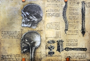
Background imageCranium Collection: Anatomical Drawings of Two Skulls in Profile to the Left, c1480 (1945). Artist: Leonardo da Vinci

Anatomical Drawings of Two Skulls in Profile to the Left, c1480 (1945). Artist: Leonardo da Vinci
Anatomical Drawings of Two Skulls in Profile to the Left, c1480 (1945). From The Drawings of Leonardo da Vinci. [Reynal & Hitchcock, New York, 1945]

Background imageCranium Collection: Ancient Cimbrian, Esthonian Finn, Lappe, 1848

Ancient Cimbrian, Esthonian Finn, Lappe, 1848. An engraving from the Natural History of Man, by James Cowles Prichard, (Hippolyte Bailliere, London, third edition, 1848)

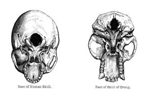
Background imageCranium Collection: Human and orang-utan skulls, 1848

Human and orang-utan skulls, 1848. Illustrations of the base of the skull. An engraving from the Natural History of Man, by James Cowles Prichard, (Hippolyte Bailliere, London, third edition, 1848)

Background imageCranium Collection: Self-portrait, c1850-1867. Artist: Charles Pierre Baudelaire

Self-portrait, c1850-1867. Artist: Charles Pierre Baudelaire
Self-portrait, c1850-1867. From a private collection

Background imageCranium Collection: Franz Joseph Gall, German physician and founder of Phrenology, c1790

Franz Joseph Gall, German physician and founder of Phrenology, c1790
Franz Joseph Gall, German physician and founder of Phrenology, c1820. Phrenology was a theory, propounded by Gall (1758-1828) in around 1800

Background imageCranium Collection: Phrenology, 1820 (1886). Artist: Frank Dadd

Phrenology, 1820 (1886). Artist: Frank Dadd
Phrenology, 1820 (1886). Measuring the bumps on a boys head to assess his future. On the wall in the background is a portrait of of Franz Josef Gall (1757-1828)

Background imageCranium Collection: Phrenology chart, showing presumed areas of activity of the brain, c1920

Phrenology chart, showing presumed areas of activity of the brain, c1920. Phrenology was a theory, propounded by the German physician Franz Joseph Gall in around 1800

Background imageCranium Collection: Skull of Piltdown Man (Eanothropus daswoni), 1912

Skull of Piltdown Man (Eanothropus daswoni), 1912. This supposed fossil was discovered near Lewes, Sussex, in 1912 by Charles Dawson, a local solicitor and amateur archaeologist

Background imageCranium Collection: Model of the skull of Piltdown Man (Eanothropus dawsoni), 1914. Artist: Dr Smith Woodward

Model of the skull of Piltdown Man (Eanothropus dawsoni), 1914. Artist: Dr Smith Woodward
Model of the skull of Piltdown Man (Eanothropus dawsoni), 1914. Model as reconstructed by Dr Smith Woodward. Dark areas are from the original fossil, the light are the restored areas

Background imageCranium Collection: Java Man or Pithecanthropus erectus (Homo Erectus erectus)

Java Man or Pithecanthropus erectus (Homo Erectus erectus). Early human fossills discovered on island Java (Indonesia). Paleolithic

Background imageCranium Collection: Medicine. Study of anatomy by Leonardo Da Vinci. 15th centur

Medicine. Study of anatomy by Leonardo Da Vinci. 15th centur
Study of anatomy by Leonardo Da Vinci. 15th century. Skeletal structure. National Museum of Science and Technology Leonardo da Vinci. Milan. Italy

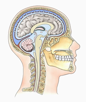
Background imageCranium Collection: Lateral cross section through the skull showing the brain with intermediate layers (meninges)

Lateral cross section through the skull showing the brain with intermediate layers (meninges) highlighted

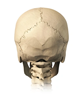
Background imageCranium Collection: Anatomy of human skull, rear view

Anatomy of human skull, rear view

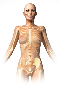
Background imageCranium Collection: Female body with bone skeleton and internal organs superimposed

Female body with bone skeleton and internal organs superimposed
Anatomy of female body with bone skeleton and all internal organs superimposed

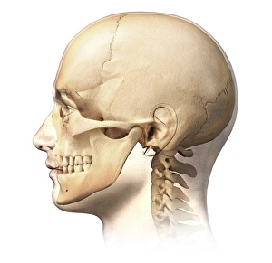
Background imageCranium Collection: Male human head with skull in ghost effect, side view

Male human head with skull in ghost effect, side view. on white background

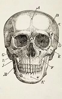
Background imageCranium Collection: Anterior view of human skull, with labels

Anterior view of human skull, with labels

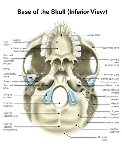
Background imageCranium Collection: Base of human skull, inferior view, with labels

Base of human skull, inferior view, with labels

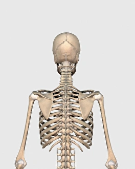
Background imageCranium Collection: Rear view of human skeletal system showing upper back

Rear view of human skeletal system showing upper back

Background imageCranium Collection: Anatomy of human skull, cutaway view with half brain showing

Anatomy of human skull, cutaway view with half brain showing, front view

Background imageCranium Collection: X-ray view of a naked woman walking, with skeletal bones superimposed

X-ray view of a naked woman walking, with skeletal bones superimposed

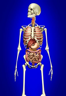
Background imageCranium Collection: Male skeleton with internal organs on blue background

Male skeleton with internal organs on blue background

Background imageCranium Collection: Male human skeleton in dynamic posture, front view

Male human skeleton in dynamic posture, front view

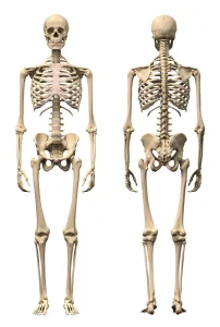
Background imageCranium Collection: Anatomy of male human skeleton, front view and back view

Anatomy of male human skeleton, front view and back view

Background imageCranium Collection: 3D rendering of a naked woman walking, with skeletal bones superimposed

3D rendering of a naked woman walking, with skeletal bones superimposed

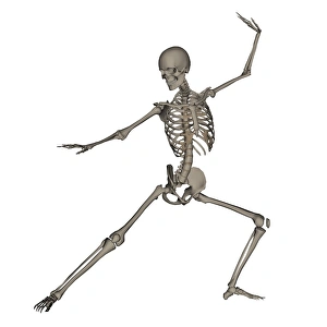
Background imageCranium Collection: Front view of human skeleton in fighting stance

Front view of human skeleton in fighting stance, isolated on white background

Background imageCranium Collection: Front view of a human skeleton posing

Front view of a human skeleton posing, isolated on white background

Background imageCranium Collection: Colored base of human skull, inferior view, with labels

Colored base of human skull, inferior view, with labels

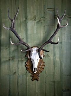
Background imageCranium Collection: Hunting trophy, 14-point-antlers, mounted red deer antlers on a wooden wall

Hunting trophy, 14-point-antlers, mounted red deer antlers on a wooden wall

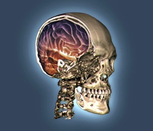
Background imageCranium Collection: Normal skull and brain, 3D CT scan C016 / 6333

Normal skull and brain, 3D CT scan C016 / 6333
Normal skull and brain. Coloured 3D computed tomography (CT) scan of the head of a 35 year old patient. Part of the skull has been cut-away to show the brain (purple and red)

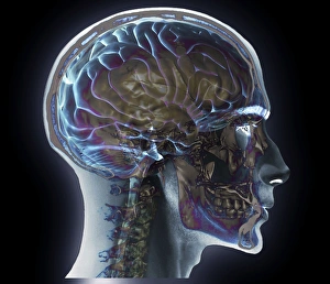
Background imageCranium Collection: Normal head and neck, MRI and 3D CT scans C016 / 6337

Normal head and neck, MRI and 3D CT scans C016 / 6337
Normal head. Coloured composite image of a magnetic resonance imaging (MRI) scan of the brain and a 3D computed tomography (CT) scan of the head and neck of a 35 year old patient

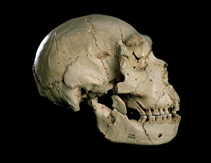
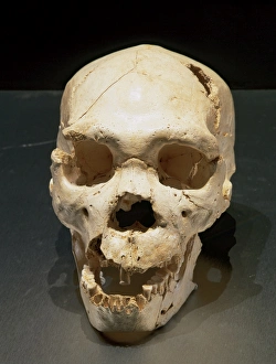
Background imageCranium Collection: Homo heidelbergensis skull (Cranium 5) C015 / 6921

Homo heidelbergensis skull (Cranium 5) C015 / 6921
Homo heidelbergensis skull (Cranium 5). Excavated in 1992 from the Sima de los Huesos pit in the Atapuerca foothills in Spain, this fossil skull dates from around 400, 000 years ago

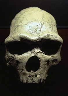
Background imageCranium Collection: Homo erectus cranium casts Peking Man & OH9

Homo erectus cranium casts Peking Man & OH9
Left: This skull is a reconstruction based upon evidence discovered at locality 1, Zhoukoudian Cave which date back 500, 000 years

Background imageCranium Collection: Homo erectus (or Homo ergaster), Turkana boy (KNM-WT 15000)

Homo erectus (or Homo ergaster), Turkana boy (KNM-WT 15000)
A replica of the fossil cranium (with reconstruction) that once belonged to a male Homo erectus aged 9 to 12 years old when he died

Background imageCranium Collection: Homo erectus, Java Man cranium (Sangiran 17)

Homo erectus, Java Man cranium (Sangiran 17)

Background imageCranium Collection: Big cat skull

Big cat skull. Side (lateral) view of the skull of a big cat with its jaws wide open. The big cats, which include tigers and lions

Background imageCranium Collection: Bones Of The Head. After A 19Th Century Print

Bones Of The Head. After A 19Th Century Print

Background imageCranium Collection: The Human Skull. From The Household Physician, Published Circa 1890

The Human Skull. From The Household Physician, Published Circa 1890

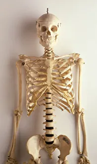
Background imageCranium Collection: Upper part of human skeleton, skull, spinal column, ribcage, shoulders, collar bones

Upper part of human skeleton, skull, spinal column, ribcage, shoulders, collar bones, upper arms and pelvis, front view

Background imageCranium Collection: Skull in profile to right, 1645. Creator: Wenceslaus Hollar

Skull in profile to right, 1645. Creator: Wenceslaus Hollar
Skull in profile to right, 1645

Background imageCranium Collection: Sectioned skull in profile to left with the left side of the cranium removed, 1651

Sectioned skull in profile to left with the left side of the cranium removed, 1651

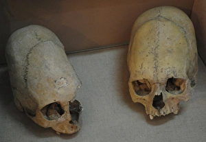
Background imageCranium Collection: Sarmatians. Deformed human skulls. Probably dated in the 3rd

Sarmatians. Deformed human skulls. Probably dated in the 3rd
Sarmatians. Two deformed human skulls. Probably dated in the 3rd century BC. Kerch Historical and Archaeological Museum. Autonomous Republic of Crimea. Ukraine

Background imageCranium Collection: Homo heidelbergensis. Skull number 5. Atapuerca, Spain

Homo heidelbergensis. Skull number 5. Atapuerca, Spain
Homo heidelbergensis. Skull number 5. Discovered in the Pit of Bones Site of Atapuerca (Spain). European Middle Pleistocene. Atapuerca. Spain

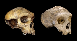
Background imageCranium Collection: Homo neanderthalensis and Homo heildebergensis

Homo neanderthalensis and Homo heildebergensis
Left: Cranium of Neanderthal man discovered at Forbes quarry Gibraltar in 1848. Right: Cranium of Broken Hill, or Rhodesian man (H. heidelbergensis) discovered at Broken Hill, Zambia in 1921

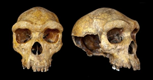
Background imageCranium Collection: Broken Hill skull, Homo heidelbergensis

Broken Hill skull, Homo heidelbergensis, discovered in Africa in 1921. The skull belonged to an adult male and may be 200, 000 to 300, 000 years old

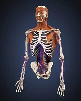
Background imageCranium Collection: Human upper body showing bones, muscles and circulatory system

Human upper body showing bones, muscles and circulatory system


All Professionally Made to Order for Quick Shipping

"Cranium: Unveiling the Mysteries of the Human Skull and Beyond" Delving into the depths of human anatomy, Leonardo da Vinci's intricate sketches of skull anatomy offer a glimpse into his fascination with unraveling the secrets held within our craniums. From his detailed drawings to modern-day advancements, we continue to explore this enigmatic structure. Hominid crania provide us with a window into our evolutionary past, showcasing how our ancestors' skulls have evolved over time. With full-body scans and MRI technology, scientists can now peer inside these ancient relics, uncovering clues about their lifestyles and adaptations. In a satirical twist on phrenology - an outdated pseudoscience that claimed personality traits could be determined by skull shape - we find humor in our quest for understanding. Yet amidst the satire lies an appreciation for how far we've come in deciphering the complexities of cranial morphology. The Paranthropus boisei (Zinjanthropus) cranium (OH5) takes us back millions of years, offering insights into early hominids' robust features. Meanwhile, X-ray images reveal intricate details of both human and animal skulls – from horses to primates – highlighting similarities and differences across species. One cannot ignore Sahelanthropus tchadensis; its fossilized skull provides a crucial link between apes and humans. This remarkable discovery challenges previous notions about our origins while fueling further exploration into humanity's beginnings. But it is not just scientific curiosity that draws attention to the cranium; it also holds significance in everyday life. Headaches become tangible through X-ray artwork, capturing both pain and beauty simultaneously. The human skull itself serves as a reminder of mortality but also showcases resilience throughout history. Finally, Homo erectus' Java Man cranium (Sangiran 17) cast allows us to step back in time once more – witnessing glimpses of ancient lives and pondering the mysteries that still surround our own existence.