
Colored Micrograph Collection
Explore the microscopic world through these captivating coloured SEMs

All Professionally Made to Order for Quick Shipping
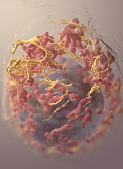
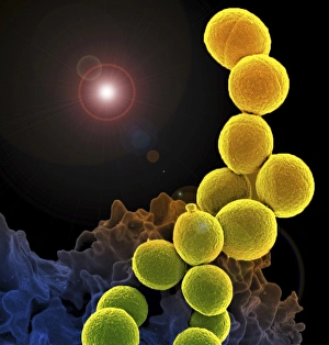
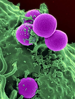
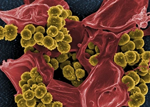
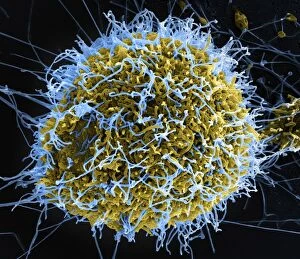
Explore the microscopic world through these captivating coloured SEMs. Witness the intricate structures of a Saturniidae moth's antennae (1), the delicate surface of a silk moth's compound eye (not shown), the complex exoskeleton of a planthopper (Hemiptera: Fulgoridae) (2), and the ominous 3D structure of a melanoma cell (3). Delve deeper into the human body with colorized SEMs of a white blood cell engulfing MRSA (4), HIV particles infecting a human T cell (5), a human neutrophil consuming MRSA (6), and Staphylococcus bacteria on a dead human neutrophil (7). Lastly, behold the ominous filamentous Ebola virus particles (8) and the surface of an HIV-infected microphage (9), offering a glimpse into the microscopic battles that occur within our bodies.