
Collenchyma Collection
Collenchyma: The Supportive Tissue in Plant Anatomy Lime tree stem, water lily leaf, beech tree leaf - what do they all have in common


All Professionally Made to Order for Quick Shipping
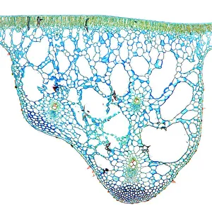
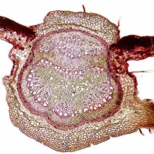
Collenchyma: The Supportive Tissue in Plant Anatomy Lime tree stem, water lily leaf, beech tree leaf - what do they all have in common? They are all plant structures that contain an important tissue called collenchyma. Under the lens of a light micrograph, this specialized tissue reveals its unique characteristics. In the lime tree stem, collenchyma cells can be seen arranged in elongated strands running parallel to each other. These cells possess thickened cell walls which provide strength and support to the growing plant. Similarly, the water lily leaf showcases collenchyma's role in maintaining rigidity as it forms a framework along its veins. Moving on to another lime tree stem micrograph, we observe how collenchyma adapts to different parts of the plant. Here, it forms irregularly shaped patches rather than distinct strands like before. This flexibility allows for efficient growth and expansion while still providing structural reinforcement. Beech tree leaves also rely on collenchyma for their resilience against environmental stressors. Microscopic examination exposes intricate patterns where these supportive cells align themselves along the leaf's surface and veins. White bryony stems exhibit yet another variation arrangement under magnification. In this case, elongated columns form a lattice-like structure within the stem walls – reinforcing them against bending or breaking forces. Sage stems display clusters of densely packed collenchymatous cells that reinforce their overall structure while allowing flexibility during wind-induced movements or physical stresses. Sunflower stems showcase both uniform and scattered arrangements of these vital tissues when observed closely through a light microscope lens. Such diversity highlights nature's adaptability even within a single species. Examining sycamore leaf veins reveals how delicate networks of interconnected collenchymatous cells serve as conduits for nutrient transport throughout plants' foliage systems – ensuring optimal growth and vitality.