
Cerebellum Collection (page 2)
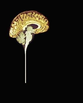
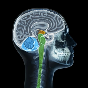
The cerebellum, often referred to as the "little brain, " is a remarkable structure located at the base of the brain


All Professionally Made to Order for Quick Shipping
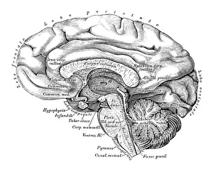
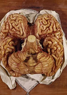
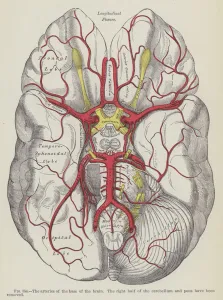
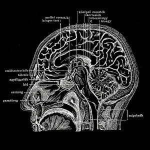
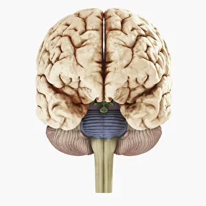
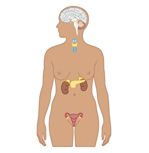
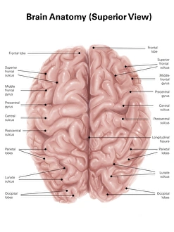
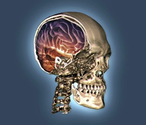
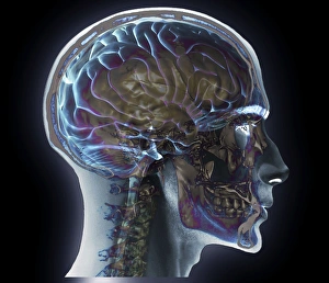
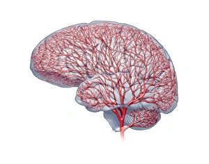
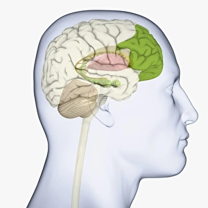
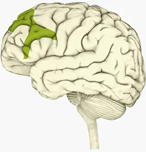
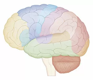
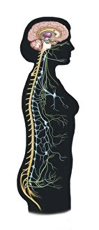
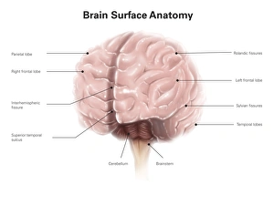
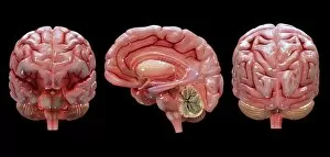
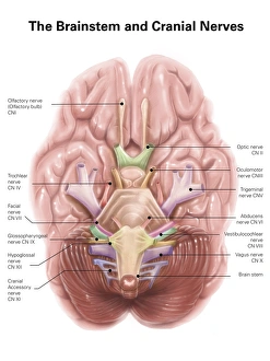
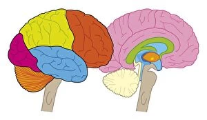
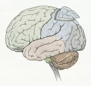
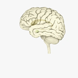
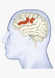
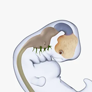
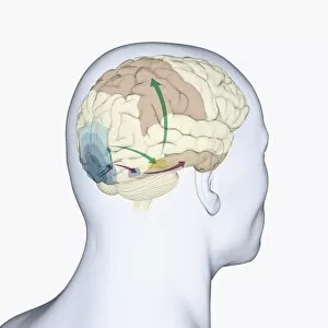
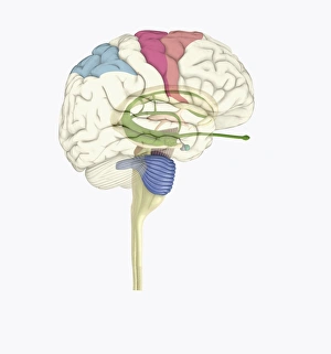
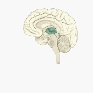
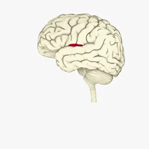
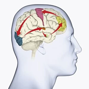
The cerebellum, often referred to as the "little brain, " is a remarkable structure located at the base of the brain. This intricate tissue plays a crucial role in coordinating movement, balance, and posture. When examined under a light micrograph, the cerebellum reveals its complex network of cells and fibers that work together seamlessly. From an inferior view of the human brain anatomy model, one can appreciate how the cerebellum fits snugly beneath the cerebral hemispheres. Its distinct lobes are clearly labeled and color-coded in a superior view of the brain. This lateral perspective allows us to understand its position relative to other regions within this vital organ. Intriguingly, Spanish histologist Santiago Ramon y Cajal's 1894 drawing showcases various cell types found within mammalian cerebellum tissue. His meticulous illustrations provide valuable insights into our understanding of this fascinating part of our brains. Glial cells are another essential component highlighted in confocal light micrographs. These support cells play a critical role in maintaining neuronal health and function within the cerebellum. Purkinje nerve cells stand out prominently when examining both artwork depicting them and actual images from MRI scans or light microscopy. These specialized neurons serve as key players in transmitting signals throughout this region of our brains. Artworks showcasing different aspects of brain anatomy further emphasize just how interconnected all these structures are within our limbic system—the emotional center responsible for memory formation and regulation among other functions.