Fraud Blocker Skip to main content

Cells Collection (page 7)

"Exploring the Intricacies of Cells: From Cerebellum Tissue to Geranium Anther" Delving into the complexity of cells

Background imageCells Collection: Myeloblast blood cell, light micrograph

Myeloblast blood cell, light micrograph
Myeloblast blood cell. Light micrograph of blood cells, including a myeloblast, a precursor for a type of white blood cell (leucocyte). Myeloblasts differentiate into granulocytes

Background imageCells Collection: Pancreatic acinar cell

Pancreatic acinar cell. Transmission electron micrograph (TEM) of a section through an enzyme-secreting acinar cell in the human pancreas, showing part of the nucleus (round, far left)

Background imageCells Collection: Promyelocyte blood cell, light micrograph

Promyelocyte blood cell, light micrograph. This blood cell (centre) is a precursor for a type of white blood cell called a granulocyte, formed by granulopoiesis in the bone marrow

Background imageCells Collection: Chronic lymphocytic leukaemia, micrograph

Chronic lymphocytic leukaemia, micrograph
Chronic lymphocytic leukaemia. Light micrograph of blood cells sampled from a lymphatic ganglion in a case of chronic lymphocytic leukaemia, also called chronic lymphoid leukaemia (CLL)

Background imageCells Collection: Hepatocyte liver cell, TEM

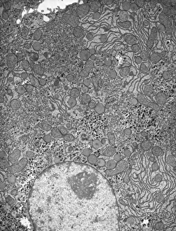
Hepatocyte liver cell, TEM
Hepatocyte liver cell. Transmission electron micrograph (TEM) of a section through an hepatocyte liver cell, showing rough and smooth endoplasmic reticulum (ER, dark lines)

Background imageCells Collection: Liver tissue, TEM

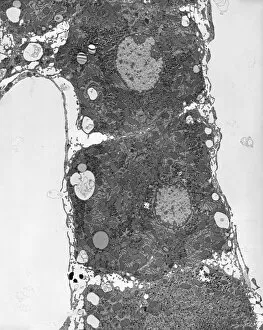
Liver tissue, TEM
Liver tissue. Transmission electron micrograph (TEM) of a section through the liver, showing part of a radial cord of hepatocyte liver cells (dark) and the vascular sinusoids (white)

Background imageCells Collection: Trachea lining, SEM C015 / 9936

Trachea lining, SEM C015 / 9936
Trachea lining. Coloured scanning electron micrograph (SEM) of a section through the lining of the trachea (wind pipe). The trachea links the larynx (voice box) to the lungs

Background imageCells Collection: Retina layers, SEM C018 / 0520

Retina layers, SEM C018 / 0520
Retina layers, coloured scanning electron micrograph (SEM). The retina of the human eye is found on the inside of the eyeball and is formed from a number of layers

Background imageCells Collection: Rod and cone cells of the eye, SEM C014 / 4864

Rod and cone cells of the eye, SEM C014 / 4864
Rod and cone cells of the eye. Coloured scanning electron micrograph (SEM) of rod and cone cells in the retina of a mammalian eye

Background imageCells Collection: Chives leaf stoma, SEM C016 / 8060

Chives leaf stoma, SEM C016 / 8060
Chives leaf stoma. Coloured scanning electron micrograph (SEM) of a stoma (oval, centre) on the surface of a leaf from a chives (Allium schoenoprasum) plant

Background imageCells Collection: Coltsfoot leaf stomata, SEM C016 / 8052

Coltsfoot leaf stomata, SEM C016 / 8052
Coltsfoot leaf stomata. Coloured scanning electron micrograph (SEM) of two stomata (round) on the surface of a leaf from a coltsfoot (Tussilago farfara) plant

Background imageCells Collection: Human olfactory system, artwork C013 / 5940

Human olfactory system, artwork C013 / 5940
Human olfactory system. Cutaway computer artwork of a persons head, with an enlargement (upper right) showing the olfactory receptor cells (red)

Background imageCells Collection: Global winds, satellite-based diagram C016 / 3731

Global winds, satellite-based diagram C016 / 3731
Global winds, satellite-based diagram. The rotation of this Earth globe (dark blue arrow) and the Coriolis effect causes the wind patterns shown

Background imageCells Collection: White blood cells and platelets, SEM C016 / 3099

White blood cells and platelets, SEM C016 / 3099
White blood cells and platelets. Coloured scanning electron micrograph (SEM) of platelets (green) with a variety of different white blood cells (leucocytes, yellow)

Background imageCells Collection: White blood cells and platelets, SEM C016 / 3098

White blood cells and platelets, SEM C016 / 3098
White blood cells and platelets. Coloured scanning electron micrograph (SEM) of platelets (purple) with a variety of different white blood cells (leucocytes, blue)

Background imageCells Collection: Red blood cells, light micrograph C016 / 3035

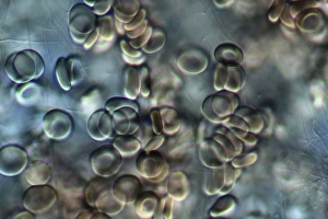
Red blood cells, light micrograph C016 / 3035
Red blood cells. Differential interference contrast (DIC) micrograph of red blood cells (erythrocytes). Red blood cells are biconcave, giving them a large surface area for gas exchange

Background imageCells Collection: Trachea lining, SEM C013 / 7126

Trachea lining, SEM C013 / 7126
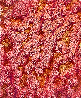
Trachea lining. Coloured scanning electron micrograph (SEM) of the lining of a trachea (wind pipe), showing the cilia (hair-like) and goblet cells (orange). The trachea links the larynx to the lungs

Background imageCells Collection: Trachea lining, SEM C013 / 7122

Trachea lining, SEM C013 / 7122
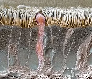
Trachea lining. Coloured scanning electron micrograph (SEM) of a section through the wall of a trachea (wind pipe). The trachea links the larynx to the lungs

Background imageCells Collection: Euglena gracilis

Euglena gracilis protists. Light micrograph of a group of Euglena gracilis protists. These organisms possess a unique combination of plant and animal characters. Like the algae, E

Background imageCells Collection: Eyelash hairs, SEM

Eyelash hairs, SEM
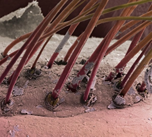
Eyelash hairs and skin. Coloured scanning electron micrograph (SEM) of eyelash hairs growing from the surface of human skin

Background imageCells Collection: Human sperm cells

Human sperm cells
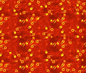
Sperm. Computer artwork of human sperm (spermatozoa), the sex cells of the male. Each sperm is about 0.065 millimetres in length

Background imageCells Collection: Bladder epithelium, light micrograph

Bladder epithelium, light micrograph
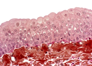
Bladder epithelium. Light micrograph of a vertical section through the wall of the urinary bladder. The inner surface is at top

Background imageCells Collection: Trachea, SEM

Trachea, SEM
Trachea. Coloured scanning electron micrograph (SEM) of the lining of the trachea (windpipe). The trachea links the larynx (voice box) to the lungs

Background imageCells Collection: Goblet cells

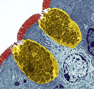
Goblet cells. Coloured transmission electron micrograph (TEM) of a section through goblet cells in the lining of the small intestine, part of the digestive tract. They are full of mucus (yellow)

Background imageCells Collection: False-colour SEM of blood cells on endocardium

False-colour SEM of blood cells on endocardium
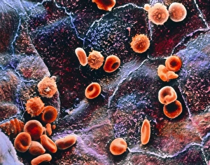
Red Blood cells inside the heart: false-colour scanning electron micrograph (SEM) of red & white blood cells on the endocardium, the lining of the heart

Background imageCells Collection: Cardiac muscle and capillary, TEM

Cardiac muscle and capillary, TEM
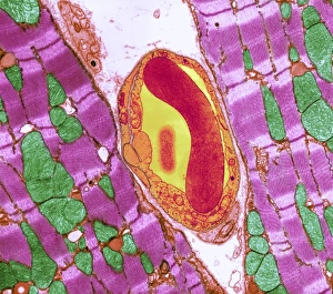
Cardiac muscle. Coloured transmission electron micrograph (TEM) of cardiac muscle fibrils (purple)from a healthy heart. Mitochondria (green) supply the muscle cells with energy

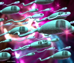
Background imageCells Collection: Human genetic engineering

Human genetic engineering. Conceptual computer artwork representing the genetic engineering of humans. Sperm cells labelled with bar codes are seen

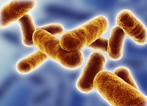
Background imageCells Collection: Rod shaped bacillus bacteria

Rod shaped bacillus bacteria
3D-computer artwork of bacillus bacteria. These rod-shaped bacteria (bacilli) are widespread in soil and in the air. Many bacillus bacteria are responsible for food spoilage

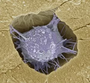
Background imageCells Collection: Osteoblast bone cell, SEM

Osteoblast bone cell, SEM
Osteoblast bone cell. Coloured scanning electron micrograph (SEM) of an osteoblast (centre) in bone tissue. Osteoblasts are primarily found in regions of new bone growth

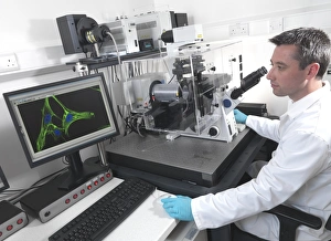
Background imageCells Collection: Cell biology laboratory

Cell biology laboratory. Scientist using confocal fluorescence microscopy to view stem cells

Background imageCells Collection: Hazel nut, light micrograph

Hazel nut, light micrograph
Hazel nut. Light micrograph of a section through a nut from a common hazel (Corylus avellana) tree, showing the large amount of fat (red) stored within endosperm tissue

Background imageCells Collection: Human ear anatomy

Human ear anatomy. Computer artwork of the structure of the human ear, showing the outer ear (left), middle ear and inner ear (right)

Background imageCells Collection: Desmosome cell junction, artwork

Desmosome cell junction, artwork
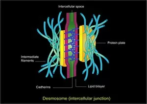
Desmosome cell junction. Computer artwork showing the structure of an adhesion junction, or desmosome. Desmosomes form the most common type of junction between epithelial cells

Background imageCells Collection: Mitochondria, SEM

Mitochondria, SEM
Mitochondria. Coloured scanning electron micrograph (SEM) of mitochondria (red) in a kidney cell. Mitochondria are a type of organelle found in the cytoplasm of eukaryotic cells

Background imageCells Collection: Skin and hair follicles, light micrograph

Skin and hair follicles, light micrograph
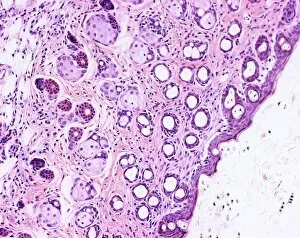
Skin and hair follicles. Light micrograph of a section through skin, showing hair follicles (dark purple circular structures). Haematoxylin and eosin stain

Background imageCells Collection: Iris root, light micrograph

Iris root, light micrograph
Iris root. Light micrograph of a section through the root of an Iris plant (Iris germanica) showing a vascular cylinder. The cylinder is comprised of a central cluster of parenchyma cells (red)

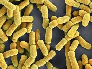
Background imageCells Collection: Lactobacillus bacteria, SEM

Lactobacillus bacteria, SEM
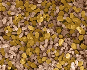
Lactobacillus sp. bacteria, coloured scanning electron micrograph (SEM). Lactobacillus bacteria are probiotic bacteria, which are useful for human and animal health as they produce bacteriocins

Background imageCells Collection: Starch grains, SEM

Starch grains, SEM
Starch grains. Coloured scanning electron micrograph (SEM) of starch grains (orange) in a black-eyed pea (Vigna sp.). Starch is synthesised from sucrose
Background imageCells Collection: Yeast cells, TEM

Yeast cells, TEM
Yeast cells, coloured transmission electron micrograph (TEM). The cell wall is shown in blue, cytoplasm in red, and vacuole in green. Yeast is a single-celled fungus that reproduces asexually

Background imageCells Collection: Midday mealtime, Wormwood Scrubs Prison, West London

Midday mealtime, Wormwood Scrubs Prison, West London
Prisoners at Wormwood Scrubs prison, west London, return to their individual cells for their midday dinner. Uniformed prison warders watch as prisoners climb the metal staircases to the galleries

Background imageCells Collection: Electricity / Daniell Cell

Electricity / Daniell Cell
DANIELL CELL Invented in 1836 by John- Frederic Daniell, English chemist and physicist, using zinc and copper

Background imageCells Collection: San Quentin Penitentiary, Marin County, California, USA

San Quentin Penitentiary, Marin County, California, USA
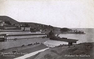
Prison cells at San Quentin Penitentiary, Marin County, California, USA. Date: circa 1910

Background imageCells Collection: Borstal New Cell Corridor

Borstal New Cell Corridor
A new-style cell corridor at an unidentified Borstal - the inmates have rooms not cells. Date: circa 1926

Background imageCells Collection: Aylesbury Inebriate Reformatory - Corridor of Cells

Aylesbury Inebriate Reformatory - Corridor of Cells
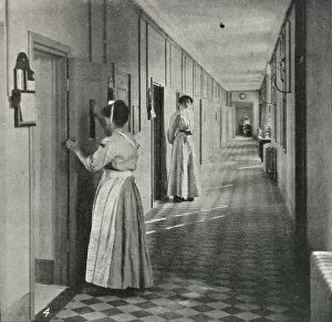
Aylesbury Inebriate Reformatory - uniformed staff on duty in a corridor of cells. Date: 1908

Background imageCells Collection: Policewoman WW1

Policewoman WW1. Captioned, You should see the Damsels (Dam-Cells) at the station. Policewoman questions nervous looking man. Date: circa 1917

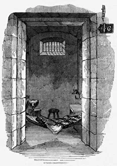
Background imageCells Collection: Pentonville prison cell interior

Pentonville prison cell interior
Pentonville prison in London: interior of cell with a hammock for sleeping. Date: 1843

Background imageCells Collection: French Prison Interior

French Prison Interior
A French prison interior, showing the guards patrolling on a gallery balcony and the doors of some of the cells. Date: early 1930s

Background imageCells Collection: Havelock Moralising

Havelock Moralising
SIR HENRY HAVELOCK Scottish military commander, urging his comrades to steer clear of drink which is the most speedy road to the guard-room and the cells. Date: 1795 - 1857


All Professionally Made to Order for Quick Shipping

"Exploring the Intricacies of Cells: From Cerebellum Tissue to Geranium Anther" Delving into the complexity of cells, we examine cerebellum tissue through a captivating light micrograph. Behind the walls of Wandsworth Prison in southwest London, cells silently carry out their vital functions within inmates' bodies. Witnessing the intricate network of nerve and glial cells, a mesmerizing light micrograph reveals their interconnectedness. At the synapse nerve junction, captured by TEM imaging, we witness the fascinating communication between cells that allows our body to function seamlessly. Within Pentonville Prison in Islington, North London, hidden stories unfold as various cell types coexist amidst confinement. Shedding light on groundbreaking research, T lymphocytes and cancer they can magnified under SEM imaging - offering hope for future treatments (SEM C001 / 1679). Peering into hippocampus brain tissue unveils a world where countless cells work together to shape our memories and emotions. Glial cells take center stage in a confocal light micrograph - showcasing their crucial role in supporting and protecting neurons within our nervous system. HeLa cells come alive under scrutiny with an intriguing light micrograph (C017 / 8299), reminding us of Henrietta Lacks' immortal contribution to medical science. Journey back in time as Aylesbury Prison's historical significance intertwines with its cellular inhabitants from 1900 onwards. Exploring beyond human boundaries, dicotyledon plant stems reveal their cellular architecture through an enchanting light micrograph. Zooming closer than ever before, geranium anthers expose their delicate beauty under SEM imaging - unveiling nature's remarkable cellular structures.