Fraud Blocker Skip to main content

Cell Biology Collection (page 4)

"Exploring the Intricacies of Cell Biology: A Fascinating Journey into Cellular Life" Nerve and glial cells, captured through a mesmerizing light micrograph

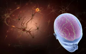
Background imageCell Biology Collection: Conceptual image of human brain with neurons

Conceptual image of human brain with neurons in background

Background imageCell Biology Collection: Red blood cells with leukocytes

Red blood cells with leukocytes

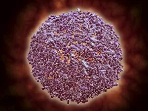
Background imageCell Biology Collection: Cluster of HIV virus

Cluster of HIV virus. HIV is the human immunodeficiency virus that can lead to acquired immune deficiency syndrom, or AIDS
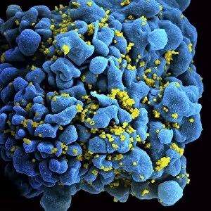
Background imageCell Biology Collection: Colorized image of HIV-infected H9 T-cell

Colorized image of HIV-infected H9 T-cell

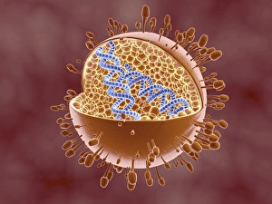
Background imageCell Biology Collection: Cutaway view of Reoviridae virus showing dna inside

Cutaway view of Reoviridae virus showing dna inside. Reoviruses can affect the gastronintestinal system and respiratory tract

Background imageCell Biology Collection: Microscopic view of ovules

Microscopic view of ovules

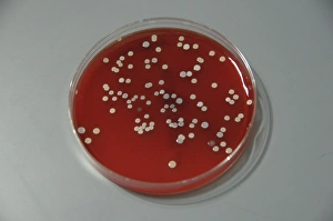
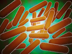
Background imageCell Biology Collection: Bacteria from human skin grown on agar in the laboratory

Bacteria from human skin grown on agar in the laboratory

Background imageCell Biology Collection: Cluster of blastomeres forming a developing morula

Cluster of blastomeres forming a developing morula (early stage of embryonic development)

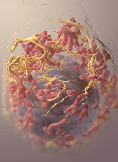
Background imageCell Biology Collection: 3D structure of melanoma cell

3D structure of melanoma cell
3D structure of a melanoma cell derived by ion abrasion scanning electron microscopy

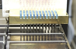
Background imageCell Biology Collection: Automated DNA sequencing laboratory equipment and paraphernalia

Automated DNA sequencing laboratory equipment and paraphernalia

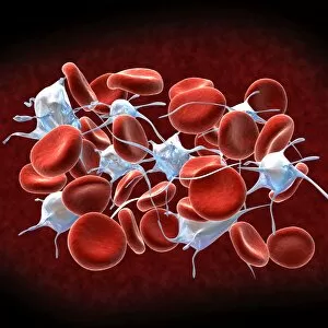
Background imageCell Biology Collection: Conceptual image of a group of platelets

Conceptual image of a group of platelets

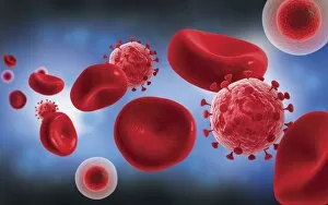
Background imageCell Biology Collection: Microscopic view of blood cells with virus

Microscopic view of blood cells with virus

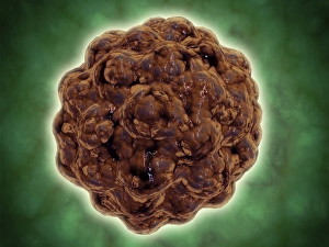
Background imageCell Biology Collection: Conceptual image of human cell

Conceptual image of human cell

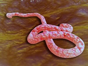
Background imageCell Biology Collection: Microscopic view of Ebola virus

Microscopic view of Ebola virus (EBOV). EBOV causes extremely severe disease in humans and in non-human primates in the form of viral hemorrhagic fever

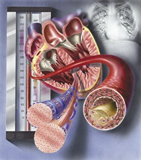
Background imageCell Biology Collection: Interior view of heart with detail of muscle cells and atherosclerotic artery

Interior view of heart with detail of muscle cells and atherosclerotic artery. Chest x-ray in background

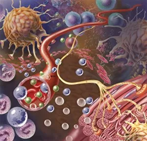
Background imageCell Biology Collection: Nerve ending, seen in lower right, sends pain message from injured muscle

Nerve ending, seen in lower right, sends pain message from injured muscle. Blood vessel and immune cells are seen in the center and upper right of image

Background imageCell Biology Collection: Thrombus forming on valve within vein

Thrombus forming on valve within vein

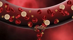
Background imageCell Biology Collection: Blood vessel with platelets, white blood cells and red blood cells

Blood vessel with platelets, white blood cells and red blood cells

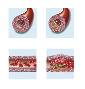
Background imageCell Biology Collection: Normal artery compared to plaque and thrombus formation in artery

Normal artery compared to plaque and thrombus formation in artery

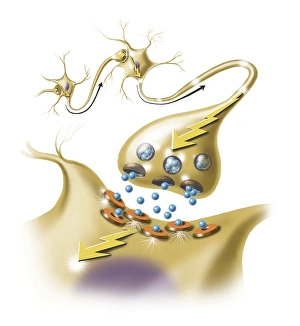
Background imageCell Biology Collection: A nerve synapse showing the release of neurotransmitters

A nerve synapse showing the release of neurotransmitters
Detail of a nerve synapse showing the release of neurotransmitters

Background imageCell Biology Collection: Conceptual image of ebola virus in artery

Conceptual image of ebola virus in artery

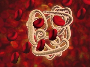
Background imageCell Biology Collection: Conceptual image of hemoglobin and red blood cells

Conceptual image of hemoglobin and red blood cells. Hemoglobin is a protein responsible for transporting oxygen in the red blood cells of vertebrates

Background imageCell Biology Collection: Microscopic view of the capsid protein of Nudaurelia capensis omega virus

Microscopic view of the capsid protein of Nudaurelia capensis omega virus

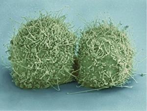
Background imageCell Biology Collection: Scanning electron micrograph of just-divided HeLa cells

Scanning electron micrograph of just-divided HeLa cells. Zeiss Merlin HR-SEM

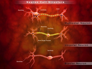
Background imageCell Biology Collection: Anatomy structure of neurons

Anatomy structure of neurons

Background imageCell Biology Collection: Conceptual image of plasmodium causing malaria

Conceptual image of plasmodium causing malaria

Background imageCell Biology Collection: Microscopic view of a stem cell

Microscopic view of a stem cell. Stem cells are biological cells found in all multicellular organisms that can divide through mitosis and differentiate into diverse specialized cell types

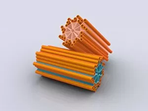
Background imageCell Biology Collection: Conceptual image of centrioles

Conceptual image of centrioles

Background imageCell Biology Collection: A blastocyst begins implanting in the wall of the uterus

A blastocyst begins implanting in the wall of the uterus

Background imageCell Biology Collection: Microscopic view of telomeres highlighted at the tips of chromosome

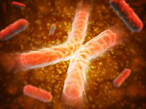
Microscopic view of telomeres highlighted at the tips of chromosome. A telomere is a region of the DNA sequence at the end of a chromosome

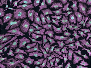
Background imageCell Biology Collection: Multiphoton fluorescence image of HeLa cells

Multiphoton fluorescence image of HeLa cells with cytoskeletal microtubules (magenta) and DNA (cyan)

Background imageCell Biology Collection: Red blood cell flow inside the artery

Red blood cell flow inside the artery

Background imageCell Biology Collection: Conceptual image of cellulose nanorods

Conceptual image of cellulose nanorods. Cellulose consists of both crystalline and amorphous regions

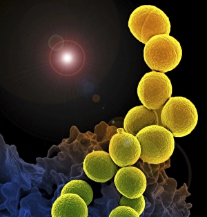
Background imageCell Biology Collection: A colorized scanning electron micrograph of a white blood cell eating MRSA

A colorized scanning electron micrograph of a white blood cell eating MRSA
A colorized scanning electron micrograph of a white blood cell eating an antibiotic resistant strain of Staphylococcus aureus bacteria, commonly known as MRSA

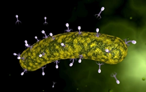
Background imageCell Biology Collection: Microscopic view of bacteriophage attacking bacteria

Microscopic view of bacteriophage attacking bacteria

Background imageCell Biology Collection: Conceptual image of synapse receptors

Conceptual image of synapse receptors

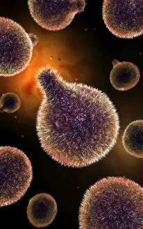
Background imageCell Biology Collection: Conceptual image of human cytomegalovirus

Conceptual image of human cytomegalovirus. Cytomegalovirus is a genus of the viral family Herpesviridae

Background imageCell Biology Collection: Microscopic view of Sindbis virus

Microscopic view of Sindbis virus (SINV). SINV is a mosquito-borne virus that causes rash and arthritis, has been causing outbreaks in humans

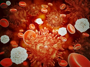
Background imageCell Biology Collection: Microscopic view of H5N1 virus with red blood cells and white blood cells

Microscopic view of H5N1 virus with red blood cells and white blood cells

Background imageCell Biology Collection: Conceptual image of male sperm

Conceptual image of male sperm

Background imageCell Biology Collection: Conceptual image of fertilization

Conceptual image of fertilization

Background imageCell Biology Collection: Conceptual image of platelets with red blood cells

Conceptual image of platelets with red blood cells
Conceptual image of platelets with white blood cells and red blood cells

Background imageCell Biology Collection: Microscopic view of Mitochondria

Microscopic view of Mitochondria. Mitochondria are the cells power producers. They convert energy into forms that are usable by the cell. Located in the cytoplasmcells activities

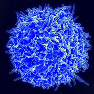
Background imageCell Biology Collection: Scanning electron micrograph of a human T cell

Scanning electron micrograph of a human T cell
Scanning electron micrograph of a human T lymphocyte (also called a T cell) from the immune system of a healthy donor

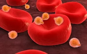
Background imageCell Biology Collection: Conceptual image of malaria parasites within red blood cells

Conceptual image of malaria parasites within red blood cells

Background imageCell Biology Collection: Conceptual image of a red blood cell

Conceptual image of a red blood cell

Background imageCell Biology Collection: Conceptual image of a platelet

Conceptual image of a platelet

Background imageCell Biology Collection: Microscopic view of Dentine

Microscopic view of Dentine. Dentine is the main supporting structure of the tooth and is the second hardest tissue in the body after enamel. It is 70% mineral and acellular


All Professionally Made to Order for Quick Shipping

"Exploring the Intricacies of Cell Biology: A Fascinating Journey into Cellular Life" Nerve and glial cells, captured through a mesmerizing light micrograph, reveal the intricate network that supports our nervous system. T lymphocytes battling cancer cells - witness this epic battle in stunning detail with SEM C001 / 1679 imagery. Anaesthetic inhibiting an ion channel - delve into the world of cellular communication as we uncover how anaesthetics affect these vital channels (C015 / 6718). Glial cells take center stage in a captivating confocal light micrograph, showcasing their crucial role in supporting neurons. Witness the beauty of glial stem cell culture under a microscope; it's like exploring an enchanting galaxy within our own bodies (light micrograph). Dendritic cells come to life through exquisite artwork, highlighting their pivotal role in immune response and defense mechanisms. Pine pollen grains unveil their intricate structure under a light microscope – nature's tiny marvels never cease to amaze us. Dive deep into the inner workings of pine stems with a captivating light micrograph – discover the building blocks that support these majestic trees. Explore the hidden wonders of lime tree stems through a mesmerizing light micrograph – witness nature's architectural masterpiece up close. Uncover various cell types brought to life through breathtaking artwork – each one playing its unique part in maintaining life's delicate balance. Immerse yourself in neural stem cell culture and witness firsthand how these remarkable cells hold immense potential for regenerative medicine. The artistic portrayal of cell membranes (C013 / 7467) invites you to ponder upon their crucial role as gatekeepers controlling molecular traffic within living organisms. Embark on this awe-inspiring journey into cell biology and unlock the secrets that shape all living beings from within.