Fraud Blocker Skip to main content

Cardiology Collection (page 2)

Cardiology, the captivating realm of the heart, unveils its intricate beauty through a myriad of hints

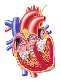
Background imageCardiology Collection: Anatomy of human heart, cross section

Anatomy of human heart, cross section with detailed internal structure

Background imageCardiology Collection: Arteries and veins of the human body

Arteries and veins of the human body

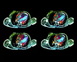
Background imageCardiology Collection: Cardiac lymphoma, MRI scans

Cardiac lymphoma, MRI scans
Brain tumour. Coloured magnetic resonance imaging (MRI) scans of axial sections through the chest of a 22-year-old patient, showing a tumorous lesion invading the right ventricle of the heart (large)
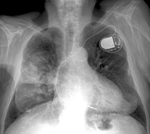
Background imageCardiology Collection: Heart and lung disease, X-ray C018 / 0498

Heart and lung disease, X-ray C018 / 0498
Heart and lung disease. Coloured X-ray of the chest of an 80 year old patient with heart and lung disease. The patient is a former miner and has chronic obstructive pulmonary disease (COPD)

Background imageCardiology Collection: Cardiac muscle, TEM

Cardiac muscle, TEM
Cardiac muscle. Coloured transmission electron micrograph (TEM) of cardiac muscle fibrils (orange)from a healthy heart. Mitochondria (pink) supply the muscle cells with energy

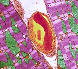
Background imageCardiology Collection: Cardiac muscle and capillary, TEM

Cardiac muscle and capillary, TEM
Cardiac muscle. Coloured transmission electron micrograph (TEM) of cardiac muscle fibrils (purple)from a healthy heart. Mitochondria (green) supply the muscle cells with energy

Background imageCardiology Collection: Cardiac stress test, artwork

Cardiac stress test, artwork
Cardiac stress test. Artwork showing a patient undergoing a cardiac stress test on a treadmill. At top are ECG (electrocardiogram) traces showing the patients heart rate at rest (left)

Background imageCardiology Collection: Pigs heart

Pigs heart. The heart of a pig is anatomically similar to that of a human. The heart is a hollow muscular organ that pumps blood around the body

Background imageCardiology Collection: Structure of a human heart, artwork

Structure of a human heart, artwork
Structure of a human heart. Computer artwork of a heart with the right ventricle (second chamber) removed, showing the inside of the left atrium (first chamber)

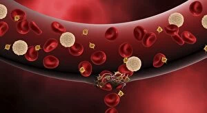
Background imageCardiology Collection: Microscopic view of blood clotting inside the artery

Microscopic view of blood clotting inside the artery

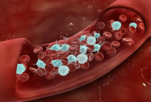
Background imageCardiology Collection: Blood vessel with platelets, white blood cells and red blood cells

Blood vessel with platelets, white blood cells and red blood cells

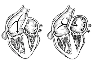
Background imageCardiology Collection: Comparison of normal heart versus heart with a patent foramen ovale

Comparison of normal heart versus heart with a patent foramen ovale

Background imageCardiology Collection: Female body sitting in dynamic posture with circulatory system superimposed

Female body sitting in dynamic posture with circulatory system superimposed
Human female body sitting in dynamic posture, with full circulatory system superimposed

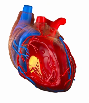
Background imageCardiology Collection: Model of a human heart

Model of a human heart

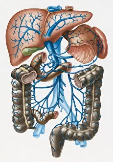
Background imageCardiology Collection: Stomach, Spleen, Liver and Intestines and Venous Drainage into Portal Vein

Stomach, Spleen, Liver and Intestines and Venous Drainage into Portal Vein

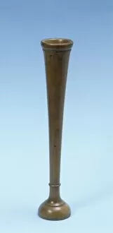
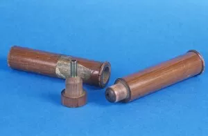
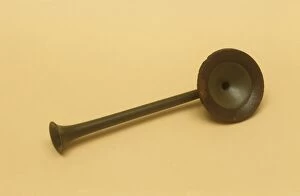
Background imageCardiology Collection: Laennec monaural stethoscope, circa 1820 C017 / 6951

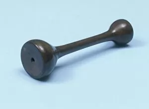
Laennec monaural stethoscope, circa 1820 C017 / 6951
English monaural stethoscope, made out of fruitwood, dating from the year 1820. French physician, Rene Theophile Hyacinthe Laennec invented the monaural stethoscope in 1816

Background imageCardiology Collection: Telemedicine, conceptual image C017 / 7607

Telemedicine, conceptual image C017 / 7607
Telemedicine, conceptual image

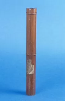
Background imageCardiology Collection: Extra long monaural stethoscope, circa 18 C017 / 6948

Extra long monaural stethoscope, circa 18 C017 / 6948
Unusual extra long monaural stethoscope dating from 1860. This piece was made in England out of fruitwood. French physician

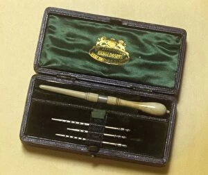
Background imageCardiology Collection: Drainage trocar with cannulae, circa 1860 C017 / 6949

Drainage trocar with cannulae, circa 1860 C017 / 6949
Dr. Southeys drainage trocar with 3 cannulae in their case. This set dates from 1860 and is made by Arnold and Sons, England

Background imageCardiology Collection: Telemedicine, conceptual image C017 / 7606

Telemedicine, conceptual image C017 / 7606
Telemedicine, conceptual image

Background imageCardiology Collection: Medical care, conceptual artwork F006 / 3979

Medical care, conceptual artwork F006 / 3979
Medical care, conceptual computer artwork

Background imageCardiology Collection: Mechanical heart, conceptual artwork F006 / 3720

Mechanical heart, conceptual artwork F006 / 3720
Mechanical heart, conceptual computer artwork

Background imageCardiology Collection: Heart health, conceptual artwork F006 / 3776

Heart health, conceptual artwork F006 / 3776
Heart health, conceptual computer artwork

Background imageCardiology Collection: ECG, artwork F006 / 4606

ECG, artwork F006 / 4606
ECG. Computer artwork of an electrocardiogram (ECG) showing a normal heart rate

Background imageCardiology Collection: Mechanical heart, conceptual artwork F006 / 3721

Mechanical heart, conceptual artwork F006 / 3721
Mechanical heart, conceptual computer artwork

Background imageCardiology Collection: Tablet computer, heart attack artwork F006 / 4625

Tablet computer, heart attack artwork F006 / 4625
Tablet computer showing conceptual artwork of a heart attack

Background imageCardiology Collection: Tablet computer, heart attack artwork F006 / 4628

Tablet computer, heart attack artwork F006 / 4628
Tablet computer showing conceptual artwork of a heart attack

Background imageCardiology Collection: ECG trace and head with brain, artwork

ECG trace and head with brain, artwork
ECG trace and brain. Computer artwork of an ECG (electrocardiogram) trace in front of a head and a human brain. An ECG uses electrodes to measure the electrical activity of the heart during

Background imageCardiology Collection: ECG, computer artwork

ECG, computer artwork
ECG. Computer artwork of an electrocardiogram (ECG) showing a normal heart rate

Background imageCardiology Collection: ECG trace and mri brain scan, artwork

ECG trace and mri brain scan, artwork
Brain anatomy. Coloured magnetic resonance imaging (MRI) scan of the human head from the side. The sagittal scan has halved the brain, revealing the internal anatomy

Background imageCardiology Collection: Heartbeat trace, artwork C018 / 8585

Heartbeat trace, artwork C018 / 8585
Heartbeat trace. Artwork of the output trace from an electrocardiograph (ECG), a method used to monitor the beating of the heart

Background imageCardiology Collection: Heartbeat alert, artwork C018 / 8588

Heartbeat alert, artwork C018 / 8588
Heartbeat alert. Artwork of world alert with the output trace from an electrocardiograph (ECG), a method used to monitor the beating of the heart

Background imageCardiology Collection: Heartbeat trace, artwork C018 / 8586

Heartbeat trace, artwork C018 / 8586
Heartbeat trace. Artwork of the output trace from an electrocardiograph (ECG), a method used to monitor the beating of the heart

Background imageCardiology Collection: Heartbeat trace, artwork C018 / 8587

Heartbeat trace, artwork C018 / 8587
Heartbeat trace. Artwork of the output trace from an electrocardiograph (ECG), a method used to monitor the beating of the heart

Background imageCardiology Collection: Physical fitness test C014 / 1103

Physical fitness test C014 / 1103
Physical fitness test. Athlete riding an exercise bike while his oxygen consumption is measured with a face mask. Electrodes on his chest monitor his heart rate

Background imageCardiology Collection: Physical fitness test C014 / 1108

Physical fitness test C014 / 1108
Physical fitness test. Athlete riding an exercise bike while his oxygen consumption is measured with a face mask. Electrodes on his chest monitor his heart rate (shown on screen)

Background imageCardiology Collection: Heart-lung anatomy, 1825 artwork

Heart-lung anatomy, 1825 artwork
Heart-lung anatomy. Axial section through the chest at the level of the fifth thoracic vertebra (T5). The front of the body is at top

Background imageCardiology Collection: Monaural stethoscope, circa 1870 C017 / 6975

Monaural stethoscope, circa 1870 C017 / 6975
Monaural stethoscope dating from 1870. The stethoscope is made of gutta percha with a rubber ring at the earpiece end which also functions as a percussor

Background imageCardiology Collection: Monaural stethoscope, circa 1870 C017 / 6937

Monaural stethoscope, circa 1870 C017 / 6937
Billings type monaural stethoscope, made out of fruitwood, dating from around 1870, England. French physician, Rene Theophile Hyacinthe Laennec invented the monaural stethoscope in 1816

Background imageCardiology Collection: Telemedicine, conceptual image C017 / 7589

Telemedicine, conceptual image C017 / 7589
Telemedicine, conceptual image

Background imageCardiology Collection: Monaural stethoscope, circa 1820 C017 / 6930

Monaural stethoscope, circa 1820 C017 / 6930
English monaural stethoscope, made out of fruitwood, dating from the year 1820. French physician, Rene Theophile Hyacinthe Laennec invented the monaural stethoscope in 1816

Background imageCardiology Collection: Physical fitness test C014 / 1105

Physical fitness test C014 / 1105
Physical fitness test. Athlete riding an exercise bike while his oxygen consumption is measured with a face mask. Electrodes on his chest monitor his heart rate

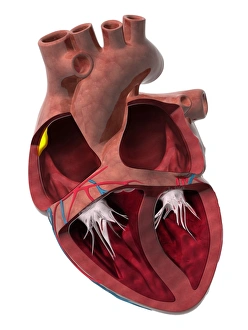
Background imageCardiology Collection: Heart chambers and sinus node, artwork C014 / 2030

Heart chambers and sinus node, artwork C014 / 2030
Heart chambers and sinus node (yellow). Cutaway artwork revealing the four internal heart chambers and the heart valves and tendons (white). The two upper chambers are the atria

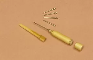
Background imageCardiology Collection: Drainage trocar with cannulae, circa 1860 C017 / 6950

Drainage trocar with cannulae, circa 1860 C017 / 6950
Dr. Southeys drainage trocar with 3 cannulae arranged on an orange background. This set dates from 1860 and is made by Arnold and Sons, England. The instruments are made from ivory and silver

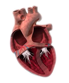
Background imageCardiology Collection: Heart chamber anatomy, artwork C014 / 2029

Heart chamber anatomy, artwork C014 / 2029
Heart chamber anatomy. Cutaway artwork revealing the four internal heart chambers and the heart valves and tendons (white). The two upper chambers are the atria

Background imageCardiology Collection: Telemedicine, conceptual image C017 / 7588

Telemedicine, conceptual image C017 / 7588
Telemedicine, conceptual image

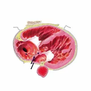
Background imageCardiology Collection: Atrial extrasystole, anatomical artwork

Atrial extrasystole, anatomical artwork
Atrial extrasystole. Anatomical computer artwork of a transverse section through a human heart during atrial contraction in extrasystole

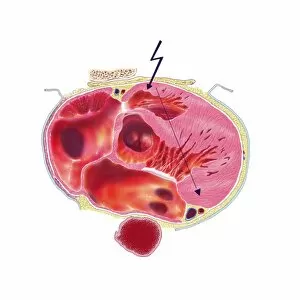
Background imageCardiology Collection: Ventricular extrasystole, anatomical artw

Ventricular extrasystole, anatomical artw
Atrial extrasystole. Anatomical computer artwork of a transverse section through a human heart during ventricular contraction in extrasystole


All Professionally Made to Order for Quick Shipping

Cardiology, the captivating realm of the heart, unveils its intricate beauty through a myriad of hints. From ECGs showcasing the rhythmic dance of a normal heart rate to mesmerizing artwork depicting the human heart's anatomy, this field unravels the secrets held within our most vital organ. In awe-inspiring illustrations, artists bring to life the complexity and elegance of the human heart. Their brushstrokes breathe life into each chamber and vessel, while their colors mirror its vibrant energy. The harmonious connection between our hearts and lungs is depicted in breathtaking images that celebrate their symbiotic relationship. A chest X-ray reveals a healthy human heart, beating with strength and vigor—a testament to its resilience. Even in space, where gravity loses its grip on earthly matters, the heart remains an enigma; an ethereal image floating amidst celestial wonders. Resin casts capture every delicate detail of intricate blood vessels that intricately weave throughout this remarkable organ. These lifelines carry crimson red blood cells on their journey through arteries and veins—nourishing every corner of our being. Yet not all hearts are untouched by affliction; some bear witness to disease's cruel touch. In haunting depictions, we confront these realities head-on—the weakened muscle struggling against adversity—an urgent reminder for us to cherish our own health. Delving into history's annals takes us back centuries ago when anatomists meticulously studied hearts with fervor unmatched. Their discoveries paved pathways towards understanding cardiac physiology—a legacy still cherished today. Cardiology encompasses more than just medical expertise—it intertwines artistry with science as it seeks answers about one of humanity's greatest mysteries: our own heartbeat. It reminds us that within each pulsating rhythm lies a story waiting to be told—a tale written by countless generations who have marveled at this wondrous symbol of life itself.